
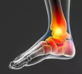
CALCANEAL NAVICULAR COALITION
The calcaneal navicular coalition refers to a congenital condition in which the calcaneus (heel bone) and navicular (midfoot bone) become fused by bone or fibrous tissue, resulting in symptoms such as pain, stiffness, and difficulty walking. This condition is one of the specific types of tarsal coalition, which involves an abnormal connection between foot bones.
At the Medical City Children’s Orthopedics and Spine Specialists medical practice, our expert Calcaneal Navicular Coalition Doctors and Surgeons are dedicated to diagnosing and treating children with all types of medical conditions to ensure comprehensive care is tailored to each patient’s needs. We specialize in children’s health and have completed advanced training. We treat both minor and severe medical conditions with compassion, and our team treats children with irregular foot conditions.
If your child needs surgery or casting, our Fracture Care Clinic opens every day and you do not need an appointment. Surgery rooms get scheduled every morning, so your child receives the care and attention they need right away.
What Causes Calcaneal Navicular Coalition
The calcaneal navicular coalition refers to a congenital (present at birth) condition. This means that the condition occurs during the development of the foot bones while the fetus grows in the womb. Doctors and researchers do not fully understand the cause of this abnormality, but scientists believe genetics and other factors affect this condition.
Understanding Tarsal Coalition
Tarsal coalition generally manifests in late childhood or early adolescence, limiting foot movement and causing discomfort. An abnormal connection can occur between various bones in the midsection and back of the foot. This can affect mobility and increase the likelihood of frequent ankle sprains. The condition comprises bone, cartilage, or fibrous tissue and occurs in both feet in about half of all cases.
Understanding Accessory Navicular and Its Symptoms
An accessory navicular is an additional bone growth found near the navicular bone, sitting inside the posterior tibial tendon. This condition is present from birth and often results from an incomplete fusion between the extra bone and the navicular during the bone’s developmental phase.
Common Symptoms
- Pain and Tenderness: Individuals with accessory navicular often experience discomfort, primarily manifesting as pain and tenderness in the affected area.
- Positioned on the Inner Foot: The pain usually occurs on the inner side of the foot. Especially noticeable when engaging in activities that put pressure on this region.
- Irritation During Movement: The abnormal movement between the accessory bone and the navicular can exacerbate the pain, leading to increased discomfort over time.
QUESTIONS AND ANSWERS
Define Calcaneal Navicular Coalition?
Calcaneal Navicular Coalition refers to a congenital condition where the bones of the calcaneus (heel bone) and navicular (foot bone) fuse together
Describe the symptoms of Calcaneal Navicular Coalition?
Symptoms of Calcaneal Navicular Coalition include pain in the foot and ankle, difficulty walking, and limited range of motion in the foot.
How do doctors treat Calcaneal Navicular Coalition?
Treatment for Calcaneal Navicular Coalition depends on the severity of the condition and may include non-surgical options such as physical therapy, orthotics, and pain management, or surgical options such as arthrodesis (fusion) or osteotomy (bone cutting). Doctors treat this condition to relieve pain and improve function.
The Medical City Children’s Orthopedics and Spine Specialists are experts when it comes to children and their health. To get the very best for your child, make an appointment with a children’s specialist
The Symptoms of Calcaneal Navicular Coalition can vary, but may include:
- Pain: Pain in the foot, especially in the midfoot, appears as the most common symptom of Calcaneal navicular coalition.
- Swelling: Swelling in the midfoot may also occur.
- Stiffness: The affected foot may feel stiff and have a limited range of motion.
- Foot deformity: In some cases, the affected foot may appear deformed, with an abnormally high arch or other changes in the shape of the foot.
- Difficulty walking: Pain and stiffness can make it difficult to walk, and may lead to a limp or other changes in gait.
Symptoms occur when patients walk on uneven surfaces like sand or gravel, requiring constant foot adjustment. It’s important to recognize that symptoms might not appear until later in life, and the condition might not be diagnosed until adulthood. Early diagnosis and treatment can help prevent the condition from worsening and reduce the risk of complications.
Diagnoses
Doctors diagnose the calcaneal navicular coalition by a combination of physical examination, medical history, and imaging tests. The following tests diagnose this condition:
- Physical examination: This includes a thorough examination of the foot. It includes a range of motion testing and evaluation of any tenderness or swelling in the affected area.
- X-rays: X-rays of the foot can identify the bones and detect any abnormalities, such as the fusion of the calcaneus and navicular bones.
- MRI: Magnetic Resonance Imaging, an advanced imaging test, provides a detailed view of the soft tissues and bones in the foot. This scan can confirm a diagnosis of the Calcaneal navicular coalition.
 In some cases, doctors order additional tests, such as a CT scan or a bone scan. These tests can obtain a more complete picture of the condition and its impact on the foot.
It is important to seek medical attention if your child is experiencing pain or discomfort in the foot. An early diagnosis and treatment can improve outcomes and help prevent the condition from becoming more severe.
Children’s Foot Deformities
Pediatric foot deformities encompass a variety of conditions impacting the structure and function of a child’s foot. These conditions often involve the bones, tendons, or muscles, potentially affecting how a child walks or stands.
Commonly addressed pediatric foot deformities include:
- Cavus Foot: Characterized by a high arch, this condition can lead to instability and discomfort.
- Tarsal Coalition: Involves abnormal connections between bones, causing stiffness and reduced foot movement.
- Clubfoot: A congenital condition where the foot appears twisted out of shape or position.
- Accessory Navicular: An extra bone or piece of cartilage located on the inner side of the foot just above the arch.
- Juvenile Bunion: A bony bump on the joint at the base of the big toe, often observed in adolescents.
These conditions, frequently treated by pediatric specialists, restore proper foot function and improve quality of life.
Treatment Approaches
Initial treatment strategies are typically non-surgical, aiming to alleviate symptoms through conservative methods:
- Orthotics: Special shoe inserts provide support and reduce strain on the foot.
- Casting: Doctors may recommend a short period of casting to allow the foot to rest and reduce inflammation.
For persistent pain, surgical intervention involves removing the additional bone. This procedure is generally straightforward, requiring minimal recovery time and usually resulting in significant symptom relief.
How Pediatric Foot Deformity Treatments Differ from Adult Treatments
Treating foot deformities in children requires a unique approach compared to adults. There are distinct techniques designed to cater specifically to the needs of younger patients.
Growth Plates: A Key Focus
Children’s feet continue to grow, which means it’s crucial to preserve the integrity of the growth plates. Unlike adult treatments, which may involve more straightforward corrective measures, pediatric treatments must ensure that the foot can continue developing naturally.
Specialized Techniques
Pediatric orthopedists employ a mix of non-operative and operative methods tailored for children. These might include:
- Non-Operative Interventions: Bracing, physical therapy, and custom orthotics can assist foot development without surgery.
- Operative Techniques: When surgery is necessary, procedures minimize impact on growth and function.
A Holistic Approach
The goal is to support not just the immediate correction of the deformity but also the long-term development of a healthy, functional foot. This child-centered approach ensures that treatments are not only effective in the short term but also beneficial as the child continues to grow.
How Is Juvenile Bunion Treated, and When Is Surgery Considered?
Treating juvenile bunions typically begins with non-surgical methods to manage the condition and alleviate discomfort. These conservative approaches prevent further issues and delay or eliminate the need for surgical intervention.
Non-Surgical Approaches
- Footwear Adjustments: Encouraging the use of sneakers or shoes with a wide toe box can minimize pressure on the bunion. It is advisable to avoid narrow dress shoes and high heels, which can exacerbate the symptoms.
- Activity Modifications: Sometimes, limiting activities that cause pain and discomfort can help. This might include reducing participation in activities that put undue stress on the feet.
For many young patients, these non-operative treatments are sufficient. They can significantly reduce pain and halt the progression of the bunion, diminishing the need for more invasive solutions.
Diagnosing and Treating Cavus Foot in Children
Understanding Cavus Foot Diagnosis
Cavus foot, characterized by an abnormally high arch, often involves both feet and can worsen over time. Diagnosing this condition involves careful observation of the foot’s structure and function. Doctors look for indicators such as pain, calluses, or unusual foot positioning, like the heel turning inward — a condition known as cavovarus deformity. Early symptoms might suggest underlying neuromuscular disorders, such as Charcot-Marie-Tooth disease, a hereditary condition leading to slowed nerve conduction and muscle weakness in the extremities.
To aid diagnosis, healthcare professionals may utilize X-rays to assess bone structure and pinpoint abnormalities. Additionally, a thorough physical examination allows evaluation of symptoms and any possible neurological issues, ensuring that the cavus foot isn’t linked to more severe conditions like spinal cord abnormalities.
Approaches to Treatment
Treatment strategies for cavus foot vary based on the severity of the deformity. For mild cases, pediatric specialists often recommend using orthotics, which are shoe inserts aimed at redistributing weight more evenly across the foot. This method may help alleviate discomfort and prevent progression.
However, more pronounced deformities typically require surgical intervention to restore proper alignment. Surgical procedures may involve osteotomies—cutting and repositioning bones—as well as procedures to release tight ligaments and transfer tendons. These surgeries work to balance and support weakened muscles, providing long-term relief and functionality.
Importance of Specialist Care
Given the complex nature of cavus foot correction, children must receive treatment from pediatric orthopedic specialists. Unlike adult treatment, children’s procedures must account for ongoing growth, requiring strategies like careful placement of pins and screws to allow natural development. Each surgical plan is tailored to the child’s specific needs, considering factors such as foot appearance, X-ray results, and individual symptoms.
In conclusion, while cavus foot poses significant challenges, a combination of thorough diagnosis and customized treatment plans can offer effective management for children, improving their quality of life and mobility.
When Is Surgery Considered?
Surgical options are generally reserved for instances where non-surgical treatments do not provide sufficient relief, particularly when pain disrupts daily activities. Surgery may occur when:
- Growth Considerations: The child is nearing the end of their growth phase. This timing minimizes the risk of damage to the growth plates and the chance of recurrence.
- Significant Pain: The pain is persistent and interferes with everyday life, particularly if it limits a young patient’s ability to engage in normal activities.
- Severe Deformity: In cases where the bunion is severe, doctors may recommend surgery to realign the bone and correct the toe’s position.
The specific surgical approach depends on various factors, including the bunion’s type and severity, the child’s age, and how much growth is anticipated in the future. Each case is unique, and treatment plans are tailored to meet the individual needs of the patient, ensuring the best possible outcome.
How is Accessory Navicular Treated in Children?
Accessory navicular in children involves an extra bone that forms on the inner side of the foot, connected to the navicular bone. This condition often causes discomfort and tenderness due to the additional bony growth.
Initial Non-Surgical Treatments
When treating accessory navicular in children, the first line of approach is typically non-surgical. The following provides the most common steps taken:
- Orthotics: Custom shoe inserts often provide support and reduce stress on the affected area.
- Rest and Casting: A brief period of immobilization, like casting, may allow the foot to rest and heal.
These measures aim to alleviate pain and prevent further irritation to the bone and surrounding tissue.
Surgical Intervention
If symptoms persist and become chronic, doctors may recommend surgery:
- Bone Removal: Surgery involves removing the extra bone. This is generally a straightforward procedure with a high success rate.
- Rehabilitation: Post-surgery, children can expect a short recovery period with rehabilitation focusing on restoring normal foot function.
Overall, whether through conservative measures or surgery, treatment for accessory navicular in children can effectively reduce pain and improve quality of life.
The Surgical Treatments
Surgical treatments may include:
- Bone or joint reconstruction: This involves separating the fused bones and reconstructing the affected joint to improve mobility and reduce pain.
- Osteotomy: This involves cutting and reshaping the bone to realign the affected joint and reduce stress on the affected area.
- Arthrodesis: This is a surgical procedure in which the joint is fused to eliminate motion and relieve pain.
- Joint implant: This involves the use of a joint implant to replace the affected joint and improve mobility.
It is important to note that surgical intervention is usually only recommended if non-surgical treatments fail. For instance, rest, physical therapy, orthotics, and pain management. The type of surgical treatment that is best for an individual with a Calcaneal navicular coalition will depend on a variety of factors. Those factors include the severity of the condition, the individual’s age and overall health, and their specific symptoms and needs.
Parents should seek a pediatric orthopedic surgeon to determine the best course of action. Choosing a specialist with extensive experience is crucial when tackling pediatric foot deformities. Such experts not only possess a deep understanding of the complex anatomy involved but are also well-versed in both non-surgical and surgical techniques.
- Experience Matters: The more procedures a surgeon performs, the more his or her expertise will develop for more positive outcomes. This underscores the value of selecting someone who frequently handles these cases.
- Comprehensive Care: An experienced surgeon will explore all relevant non-operative methods before considering surgery, ensuring that your child receives the most appropriate treatment.
By opting for a highly qualified pediatric orthopedic surgeon like those at the Medical City Children’s Orthopedics and Spine Specialists, parents know that their child will receive care tailored to their specific needs, maximizing the chances of a successful outcome.
How is Clubfoot Typically Treated in Infants?
Clubfoot, a condition often present at birth, is commonly addressed with a non-surgical method known as the Ponseti technique. This treatment involves the gentle manipulation of the infant’s foot, along with the application of casts.
Key Steps in the Ponseti Technique:
- Start Early: Treatment usually begins shortly after birth when the baby’s foot structure is most adaptable.
- Weekly Manipulations and Casting: The foot is gently manipulated and then placed in a cast. This process is repeated weekly, allowing gradual correction of the foot’s alignment.
- Bracing After Casting: Once the casting phase achieves the desired positioning, a brace is worn for an extended period. This brace helps maintain the corrected alignment and prevents the condition from recurring.
When followed accurately, the Ponseti method provides a high success rate in correcting clubfoot without the need for surgery, making it a favored approach among healthcare professionals.
The Age at Which Doctors Can Surgically Correct Calcaneal Navicular Coalition
The surgical correction of Calcaneal navicular coalition varies and depends on several factors, including:
- The severity of the condition: If the condition is causing significant pain and limitations in mobility, surgery may occur at an earlier age.
- Age and overall health of the individual: Younger individuals with Calcaneal navicular coalition who are otherwise healthy may become good candidates for surgery.
- Non-surgical treatments: If non-surgical treatments, such as physical therapy and pain management, do not work, doctors recommend surgery regardless of age.
It is important to note that the best time for surgery will depend on the individual’s specific needs and situation. A thorough evaluation by an orthopedic surgeon is recommended. The doctor’s examination will determine the most appropriate course of action. In some cases, doctors may postpone the surgery until later in life. This is usually the case if the condition does not cause significant pain or limitations in mobility.
When seeking treatment for pediatric foot deformities, parents should consider the following:
- Consultation with Specialists: Look for a pediatric orthopedic surgeon with extensive experience. Specialists are not only familiar with the latest non-operative techniques but can also ensure higher success rates if surgery becomes necessary due to their extensive procedural experience.
- Comprehensive Care: Choose a facility that offers a multidisciplinary team approach. Having access to general pediatricians, anesthesiologists, and physical therapists who specialize in pediatric care can enhance the treatment process and provide holistic support.
- Treatment Options: Understand that surgery is just one of several options. Experienced practitioners can provide a range of non-operative treatments that will help, depending on the severity and specifics of the foot deformity.
By considering these factors, parents can make informed decisions that prioritize their child’s health and long-term mobility.
The Recovery Period for Surgery on Calcaneal Navicular Coalition
The recovery period for surgery varies and depends on several factors, including:
- Type of surgery: The recovery period for more complex surgical procedures to fix the condition may last longer than for a simple osteotomy.
- Age and overall health of the individual: Younger, healthier individuals may recover quickly. Older patients with underlying health conditions may take longer.
- Compliance with postoperative care: Following the recommended postoperative care, such as physical therapy and rest, can help speed up the recovery process.
In general, recovery from surgery for Calcaneal navicular coalition can take several months to a year or more. During this time, the individual may need to use crutches or a walker. The patient will also need to limit their activities to allow the affected area to heal properly. Physical therapy and other forms of rehabilitation will help during the recovery period to restore mobility and strength.
It is important to follow the recommendations of the treating orthopedic surgeon to ensure a safe and successful recovery.
Why Choose Medical City Children’s Orthopedics and Spine Specialists
Finally, our doctors at Medical City Children’s Orthopedics and Spine Specialists, with offices in Arlington, Dallas, Flower Mound, Frisco, and McKinney, TX, understand the importance of maintaining your child’s health. In addition, our doctors, surgeons, experts, and specialists possess the training, knowledge, and experience required to take care of any ankle condition that causes pain or discomfort to your child. If your child experiences any form of ankle pain, don’t hesitate to call our office to schedule an appointment at one of our five locations.
____________________
Citation: National Library of Medicine – Calcaneal Navicular Coalition
The medical content on this page has been carefully reviewed and approved for accuracy by the Medical City Children’s Orthopedics and Spine Specialists’ qualified healthcare professionals, including our board-certified physicians and Physician Assistants. Our team ensures that all information reflects the latest evidence-based practices and meets rigorous standards of medical accuracy, with oversight from our expert spine doctors to guarantee reliability for our patients.
Call 214-556-0590 to make an appointment.
Comprehensive services for children from birth through adolescence at five convenient locations: Arlington, Dallas, Flower Mound, Frisco and McKinney.