
MEDIAL PATELLOFEMORAL LIGAMENT (MPFL) RECONSTRUCTION
Medial Patellofemoral Ligament reconstruction is also known as Kneecap Reconstruction Surgery. Replacing the kneecap can occur naturally with a doctor guiding the kneecap back in place. In addition, it can occur quickly by straightening the leg, or by surgery when everything else fails.
At the Medical City Children’s Orthopedics and Spine Specialists medical practice, our expert Knee Ligament Doctors and surgeons are dedicated to diagnosing and treating children with all types of medical conditions to ensure comprehensive care is tailored to each patient’s needs. We only specialize in children’s health, and we have undergone advanced training to do so. We treat minor and very major medical conditions with a compassionate approach, and our team cares for patients suffering from torn or ruptured Knee Ligaments.
If your child needs surgery or casting, our Fracture Care Clinic opens every day and you do not need an appointment. Surgery rooms get scheduled every morning, so your child receives the care and attention they need right away.
Medial Patellofemoral Ligament (MPFL) Reconstruction
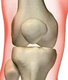
Knee Bones
A sharp blow to the knee or an unnatural twisting of the knee can lead to patellar (kneecap) dislocation. A kneecap dislocation can occur if the knee ligaments are weak or if an anatomical tendency exists. The most crucial step after a kneecap dislocation is to realign the kneecap to its correct position. This entails moving the patella into the trochlear groove. Sometimes requiring an emergency hospital visit, this sometimes occurs by simply straightening the leg.
The ligaments holding the kneecap may rip, and bits of the kneecap’s cartilage or the trochlear groove may come loose. Recurrent dislocations or partial dislocations of the patella can happen in the absence of proper treatment. In some cases, this means Kneecap Reconstruction Surgery. Also, loose tendons, ligaments, and cartilage may buckle, snag, lock, or cause additional injury.
What Factors Can Contribute to Patellar Malalignment and Instability?
Patellar malalignment is a frequent culprit in knee problems, especially among young people and athletes. Instead of gliding smoothly within the groove on the thigh bone, the kneecap may drift outside of the knee. This shift is usually toward the lateral (outer) side and can set off a cascade of issues.
Some of the main factors that contribute to this malalignment and instability include:
- Anatomic Variations: Some individuals have naturally shallow or uneven trochlear grooves, making it easier for the kneecap to slip out of place.
- Ligament Laxity: If the knee ligaments, particularly the MPFL, are loose or previously injured, the kneecap isn’t held in the center.
- Imbalanced Muscles: Weakness or imbalance in the quadriceps muscles can pull the kneecap off track.
- Maltracking of the Patella: Sometimes, the alignment of the bones in the leg results in the kneecap being pulled to the side.
- Overuse and Trauma: Repetitive activities, direct blows, or twisting injuries can strain or damage the cartilage and ligaments. This will further increase the likelihood of repeated dislocations.
Careful assessment, including a detailed history, physical exam, and imaging with X-ray, MRI, or sometimes a CT scan, is essential before planning any intervention. The underlying cause and the presence of any cartilage injury or weakness will help guide the most effective treatment plan.
Final Thoughts
Patellar dislocations can be painful, disabling, and concerning for both patients and parents. Recognizing the underlying causes, whether traumatic, anatomical, or developmental, is essential for preventing recurrence and guiding treatment. Early intervention, including strengthening exercises, bracing, and in some cases surgery, can help stabilize the knee and restore full function.
QUESTIONS AND ANSWERS
Why do children undergo kneecap reconstruction surgery?
- Recurrent Dislocations: Children who experience recurrent patellar dislocations may require surgery to stabilize the kneecap and prevent further dislocations.
- Chronic Pain: Severe patellofemoral pain that doesn’t respond to non-surgical treatments, such as physical therapy or bracing, may lead to surgery.
- Structural Abnormalities: Structural issues, such as a shallow trochlear groove (the groove in the thigh bone where the kneecap moves), can necessitate surgery to correct and realign the patella.
- Maltracking: If the patella does not track properly within the groove, it can cause pain and instability. Doctors may recommend surgery to address this problem.
How do Surgeons perform kneecap reconstruction surgery for children?
- Surgical Techniques: There are different surgical techniques used, depending on the specific issue. Common procedures include lateral release, medial patellofemoral ligament (MPFL) reconstruction, tibial tubercle transfer (e.g., TTO or Fulkerson procedure), and osteotomies to address structural abnormalities.
- Anesthesia: The surgeon will conduct the procedure under general anesthesia, and sometimes with regional nerve blocks.
- Recovery: After surgery, children may need to use crutches and wear a knee brace to support and protect the knee. Doctors will recommend Physical therapy to regain strength and range of motion.
- Activity Restrictions: Doctors will recommend certain restrictions on activities and sports for several months after surgery to allow the knee to heal.
What occurs after kneecap reconstruction surgery in children?
- Stability: The primary goal of the surgery identifies the goal to restore stability to the kneecap and prevent recurrent dislocations.
- Pain Relief: The surgery aims to alleviate chronic patellofemoral pain, allowing children to return to their normal activities with less discomfort.
- Improved Function: In many cases, the surgery results in improved knee function and range of motion.
- Long-Term Benefits: Successful surgery can have long-term benefits, reducing the risk of future knee issues.
The specific details of kneecap reconstruction surgery can vary based on the child’s age, the severity of the condition, and the surgical approach chosen by the orthopedic surgeon.
Parents and caregivers should discuss the surgery in detail with the healthcare provider, ask any specific questions they may have, and ensure that the child receives the appropriate post-operative care, including physical therapy and follow-up appointments for the best possible outcome.
It’s important to take our children to doctors who specialize in children. The doctors and surgeons at Medical City Children’s Orthopedics and Spine Specialists are experts in children and their knees.
Symptoms Indicating the Need for MPFL Reconstruction
Recurrent Patellar Dislocation or Subluxation:
-
- The child experiences repeated episodes where the kneecap fully dislocates (pops out of place) or partially shifts (subluxation) laterally, often triggered by activities like twisting, jumping, or sports (e.g., soccer or basketball, common in active communities like Coppell, TX, per your May 8, 2025, query). Dislocations may spontaneously reduce or require manual repositioning.
- Why It Matters: Recurrent dislocations (two or more episodes) indicate MPFL insufficiency, as the ligament is the primary restraint against lateral patellar movement, contributing 50–60% of stability. Chronic instability damages cartilage, increasing the risk of osteoarthritis.
- Example: A 14-year-old soccer player in Coppell reports multiple kneecap dislocations during games, with the knee “giving way” when pivoting.
Persistent Knee Pain and Swelling:
-
- Description: The child experiences ongoing pain around the front of the knee, particularly on the medial (inner) side, accompanied by swelling after activity or dislocation events. Pain may worsen with climbing stairs, squatting, or prolonged sitting (theater sign).
- Why It Matters: Pain and swelling suggest soft tissue damage, including MPFL tears or associated injuries like chondral (cartilage) defects. Persistent symptoms despite rest or therapy signal the need for surgical stabilization.
- Example: A 12-year-old complains of knee pain and swelling after dance class, unrelieved by ice or rest, indicating potential MPFL dysfunction.
Feeling of Knee Instability or “Giving Way”:
-
- Description: The child reports a sensation that the knee is unstable or about to give out, especially during weight-bearing activities like running or cutting motions. This may lead to hesitation or reduced participation in sports or play.
- Why It Matters: Instability reflects MPFL laxity or rupture, compromising patellar tracking. Recurrent instability increases the risk of further dislocations and joint damage, necessitating reconstruction to restore function.
- Example: A 15-year-old basketball player avoids jumping because their knee feels “wobbly,” a sign of chronic patellar instability.
Limited Range of Motion or Knee Stiffness:
-
- Description: The child has difficulty fully bending or straightening the knee, often due to pain, swelling, or apprehension about dislocation. Stiffness may follow acute injuries or persist with chronic instability.
- Why It Matters: Restricted motion indicates intra-articular damage (e.g., loose bodies or cartilage injury) or compensatory muscle guarding, often linked to MPFL tears. Persistent limitation despite physical therapy suggests that surgical intervention is needed.
- Example: A 13-year-old gymnast struggles to fully extend their knee after a fall, with stiffness persisting after weeks of therapy.
Visible or Palpable Patellar Maltracking:
-
- Description: The kneecap appears misaligned or moves abnormally during knee flexion, sometimes detectable by a parent or coach. The child may have a positive “J-sign” (patella deviates laterally when extending the knee) or apprehension when the patella is pushed outward.
- Why It Matters: Maltracking confirms MPFL dysfunction and underlying anatomical risk factors (e.g., trochlear dysplasia or high patella), common in children with recurrent instability. Surgical reconstruction addresses these biomechanical issues.
- Example: A parent notices their 11-year-old’s kneecap shifts sideways when climbing stairs, causing discomfort and fear of dislocation.
Associated Injuries or Symptoms:
-
- Description: The child may have signs of concurrent injuries, such as bruising, hemarthrosis (blood in the joint), or cartilage damage, often seen on MRI. They may also report popping or clicking sounds in the knee during movement.
- Why It Matters: MPFL injuries often occur with osteochondral fractures or meniscal tears, especially in traumatic dislocations (noted in your juvenile fracture query, May 3, 2025). These injuries exacerbate instability and pain, supporting the need for reconstruction to prevent further damage.
- Example: A 16-year-old reports a “pop” and bruising after a skateboarding fall, with MRI revealing an MPFL tear and chondral injury.
How Doctors Diagnose the Need for MPFL Reconstruction in a Child
Diagnosing a child who may need Medial Patellofemoral Ligament (MPFL) reconstruction involves a comprehensive clinical evaluation, medical history review, physical examination, and imaging studies. This procedure is typically considered for children who experience recurrent patellar (kneecap) dislocations, particularly when conservative treatments have failed. Below is a detailed explanation of the diagnostic process:
1. Medical History
The diagnostic process begins with a thorough review of the child’s medical history. The doctor will ask questions such as:
-
How many times has the kneecap dislocated?
-
What activities trigger the dislocations?
-
Was there trauma or a specific incident leading to the first dislocation?
-
Are there symptoms of instability, such as the kneecap feeling like it’s going to slip out?
A history of recurrent patellar instability, especially if non-operative treatment has failed, raises suspicion that the MPFL may be torn or non-functional.
2. Physical Examination
The physician performs a detailed examination of both knees, focusing on signs of instability and MPFL damage:
-
Apprehension test: The doctor gently pushes the patella outward (laterally) while watching for signs of discomfort or resistance, indicating fear of dislocation.
-
Patellar tracking: Observation of how the kneecap moves as the knee bends and straightens.
-
Tenderness along the medial patella suggests MPFL injury.
Here is the continuation and completion of the diagnostic process for determining whether a child may need Medial Patellofemoral Ligament (MPFL) reconstruction:
3. Imaging Studies
Doctors use imaging to confirm the diagnosis and assess the structural anatomy of the knee:
-
X-rays: Used to evaluate bone alignment, rule out fractures, and identify anatomical risk factors like patella alta (high-riding kneecap) or trochlear dysplasia (shallow groove in the femur).
-
MRI (Magnetic Resonance Imaging): This is the gold standard for visualizing soft tissue damage, including MPFL tears, cartilage injuries, and bone bruises from recent dislocations. It also helps confirm the integrity of other ligaments and structures in the knee.
-
CT Scan (if needed): The doctor may order a CT Scan in cases requiring detailed assessment of bone alignment or when surgical planning is underway.
4. Criteria for MPFL Reconstruction in Children
MPFL reconstruction is generally considered when:
-
The child has experienced two or more patellar dislocations.
-
Imaging confirms an MPFL tear or insufficiency.
-
There are no other more significant abnormalities that would require a different surgical intervention (e.g., tibial tubercle osteotomy).
-
Non-surgical treatments such as bracing and physical therapy have failed to control instability.
5. Conclusion
A thorough clinical and imaging-based diagnosis is essential before deciding on MPFL reconstruction. It ensures that the surgery is appropriate and targeted to restore stability, prevent future dislocations, and protect joint health as the child grows. Early and accurate diagnosis also allows doctors and parents to weigh the benefits and risks of surgery versus continued conservative care.
Who Benefits from Medial Patellofemoral Ligament (MPFL) Reconstruction?
Medial Patellofemoral Ligament (MPFL) reconstruction is typically recommended for individuals whose kneecaps are prone to slipping out of place, especially when the supporting ligaments on the inside of the knee are weak or stretched. This is often the case for people who have experienced frequent kneecap dislocations, or whose anatomy allows the kneecap to shift out of alignment easily within the first 20 to 30 degrees of knee bending—the so-called “danger zone” before the kneecap naturally settles into its groove.
How the Procedure Works
During the MPFL reconstruction procedure, surgeons create a new stabilizing ligament to help guide and secure the kneecap as the knee bends. This can occur by anchoring a new ligament between the inner side of the thigh bone (femur) and the kneecap (patella), copying the original ligament’s function. Doctors can construct a new ligament by using a sterilized donor tendon (allograft) or, also, a piece of the patient’s hamstring tendon (autograft).
The goal is to restore the medial “pull” on the kneecap at the critical point in movement, gently guiding it into its groove and helping prevent future dislocations or instability. After surgery, a knee brace is often used to limit movement while the new ligament heals, followed by a dedicated program of physical therapy. Most people can expect gradual improvement over several months as the knee regains strength and stability.
Determining the Root Cause Before Treatment
Identifying the exact reasons behind kneecap misalignment is an essential first step. Children may have more than one underlying issue, such as ligament weakness, abnormal bone structure, or repeated injuries, that contribute to their condition. Without understanding all contributing factors, treatment may miss the mark, leading to ongoing problems or additional dislocations. A tailored approach, based on a thorough evaluation, gives each patient the best chance for a stable and lasting recovery.
Kneecap Reconstruction Surgery Procedure
The Anesthesiologist will begin the first step in the procedure for cap reconstruction Surgery. He will use general anesthesia to put your child to sleep by injecting a drug into a vein. Repairing the Kneecap involves creating a new ligament to compensate for the damaged ligament. One of the thigh tendons (gracilis or semitendinosus) is removed through a small incision on the anterior medial side of the tibia. A tendon about 20 cm long is removed, and a new ligament is created.
A few more small incisions are then made in the front of the knee to reconstruct the ligaments from the anterior medial edge of the patella to the medial (medial) edge of the femur. Sutures or plastic screws, inserted into tiny drill holes, will hold the MPFL in place. The procedure may occur with one single incision, but it usually only requires three minor incisions, each ranging between 2 and 4 cm. The number of incisions also depends on the need for additional surgeries, such as a tibial tubercle osteotomy. In general, the Kneecap Reconstruction Surgery takes between 60 and 90 minutes.
What is a lateral release procedure, and when do doctors recommend it?
- Lateral Release Defined: A lateral release is a surgical step sometimes used during kneecap reconstruction. In this procedure, the surgeon carefully cuts the lateral retinaculum—a tight band of tissue on the outer side of the knee. This gives the kneecap a bit more room to shift toward the middle of the groove, helping it track properly.
- Rare as a Standalone Treatment: Surgeons rarely perform a lateral release on its own. It is uncommon that a child’s kneecap problem emanates solely from a tight lateral retinaculum. More often, this tissue becomes tight because the kneecap has been sitting too far to the outer side (laterally) for some time, usually due to other underlying structural reasons.
- Typically Combined with Other Procedures: When kneecap realignment surgery is needed, surgeons can perform a lateral release alongside other correction techniques, such as MPFL reconstruction or tibial tubercle transfer. By releasing this outer tissue, the surgeon allows the kneecap to settle back into a better, more stable position in the groove.
This approach helps ensure the kneecap stays properly aligned during movement, improving stability and reducing the risk of future dislocations.
When is lateral release combined with other procedures?
Lateral release is often performed alongside other realignment surgeries when additional correction is needed. In these cases, if the kneecap is being shifted into a more accurate or central position, releasing the tight structures on the outside of the knee (like the lateral retinaculum) helps the kneecap move smoothly into place. This combined approach allows for better alignment and improved stability of the patella.
Why is isolated lateral release rarely indicated for patellar malalignment?
Isolated lateral release is not often recommended as a stand-alone solution for patellar malalignment because it addresses a symptom rather than the underlying cause. In this procedure, the surgeon releases (or cuts) the lateral retinaculum— a band of tissue on the outer side of the knee— using minimally invasive arthroscopic techniques.
While it might seem logical to release this tissue if the kneecap is sitting too far to the outside, the real culprit is rarely just a tight lateral retinaculum. More commonly, the tightness develops as a consequence of the kneecap having been malaligned for an extended period, often due to other structural or anatomical issues. Addressing only the lateral retinaculum without correcting these primary factors usually doesn’t resolve the instability or maltracking and may even lead to further complications.
Therefore, lateral release is typically reserved for specific cases and is most effective when combined with other procedures aimed at correcting the root cause of patellar malalignment.
Risks and Drawbacks of Tibial Tuberosity Osteotomy
While tibial tuberosity osteotomy provides an effective procedure to address patellar instability, it is important to know the potential risks and drawbacks that come with this surgery.
Surgical Risks
As with any operation, infection is a possible, though uncommon, complication. There is also a risk of injury to nearby nerves or blood vessels during the procedure.
Prominent Tibial Tuberosity
After surgery, some patients may notice that the area where the bone was moved becomes more prominent. This can cause discomfort, especially while kneeling. This prominence can become a permanent change, although it does not typically affect knee function.
Hardware Concerns
The procedure generally requires one or two screws to secure the bone in its new position. Sometimes, these screws can become prominent or cause tenderness. If this happens, a subsequent minor procedure may occur to remove the hardware once the bone has healed fully.
Prolonged Rehabilitation
Recovery is gradual and involves both bracing and the use of crutches for several weeks, followed by an extended period of physical therapy. Regaining full strength and function may take anywhere from three to six months, depending on each child’s healing response.
Potential for Residual Symptoms
Some children may continue to experience mild discomfort or pain, particularly if the underlying cartilage has been damaged. Rarely, ongoing swelling or persistent symptoms may require further medical attention.
As always, the doctor will discuss the risks with the parents, and every effort is made to minimize potential complications during and after the procedure.
How Tibial Tuberosity Osteotomy Is Performed and the Path to Recovery
In some cases, your surgeon may recommend a tibial tuberosity osteotomy when there is abnormal alignment in the knee—such as increased tibial rotation or a large tibial tubercle–to–trochlear groove (TTTG) distance. This procedure involves shifting the attachment point of the patellar tendon (the tibial tuberosity) toward the inside of the tibia to help better align the kneecap.
During surgery, a small incision is made on the front of the knee. The surgeon carefully detaches a section of bone (the tuberosity) along with the attached patellar tendon. The bone segment is then gently moved medially, and sometimes slightly forward as well, to improve tracking of the kneecap. Once repositioned, the tuberosity is held securely in place—usually with one or two sturdy bone screws designed for orthopedic procedures.
After the operation, your child’s knee will need a brace to support the knee, and crutches to protect the area while it heals. Typically, this protection lasts for about six weeks. Following this period, structured physiotherapy becomes crucial to restore movement and rebuild strength. Although children start improving within a few months, a full recovery of strength and endurance may take three to six months overall.
In the long run, some patients notice a slight bump at the front of the knee. This can make kneeling a bit uncomfortable. Additionally, if screw heads become bothersome, a minor procedure might occur later to remove them.
Postoperative Recovery
Doctors refer to this surgery as day surgery. When your child wakes up, the patient will find the knee bandaged. Then, after a couple of hours, the child can go home. A physical therapist will contact the parents will monitor your child, and show him or her how to use crutches. Your child can put his or her weight on the whole leg. Children need crutches for only about 3 to 5 days after surgery and primarily use them to keep the knee from collapsing while the soft tissue heals.
The primary goals of early rehab include reducing swelling. In addition, goals include compressing the knee, lifting the leg, and gently tightening the muscles to minimize muscle wasting. Patients usually complete the physical therapy in a single day.
What Should I Expect After Kneecap Reconstruction Surgery?
It is normal to have some discomfort following Kneecap Reconstruction Surgery. During the first five days, anti-inflammatory drugs will prevent any discomfort from occurring. Additionally, your child will receive additional painkillers that your child can use as advised by your doctor. Generally speaking, pain is not severe right away after Kneecap Reconstruction Surgery. This is because local anesthesia is given at the time of the procedure to lessen discomfort after surgery.
Ice your child’s knee often, 20 minutes every hour during the day, for the first several days, as this will help to minimize discomfort and inflammation.
Outcomes for Kneecap Reconstruction Surgery
After Kneecap Reconstruction Surgery, the majority of patients may resume their usual lives, including their sports. Most experts estimate that there is a 2-4% possibility of experiencing more dislocations.
Potential Risks Associated with Kneecap Reconstruction Surgery
Infection
An infection following Kneecap Reconstruction Surgery carries a 0.5% chance. Although exceedingly unlikely, an infection can happen. To lower the risk of infection, our surgeon will prescribe antibiotics before surgery. An infection of the knee joint or a mild infection of the incisions may occur. The doctor will prescribe more Antibiotics when this occurs.
The need for hospitalization and intravenous antibiotics becomes necessary if the infection affects deeper structures and is significant. In the unlikely event that this ever occurs, and it has never occurred in our practice, a procedure on the knee will take place to remove the infection if it affects the knee joint.
Blood clots
“Deep Venous Thrombosis” – Depending on the patient, there is a 2-10% chance of getting a blood clot in a vein in your child’s leg. These blood clots occur rarely and make their way to the patient’s lungs. Pulmonary embolism refers to the medical term for this condition, and it is extremely unlikely to occur. Following your child’s operation, you should call your surgeon right away if your child experiences any calf discomfort or soreness.
The calf will undergo an ultrasound scan to check for blood clots. If they exist, the doctor will prescribe medications to stop these blood clots from growing bigger. It is crucial to let your surgeon know if you have any familial risk factors for blood clot development. This will reduce the likelihood of a blood clot.
Recurrent dislocation
The chance of the Kneecap Dislocating again after medial patellofemoral ligament repair occurs about 2-4% of the time. If your child has previously had two dislocations, this percentage rises to nearly 50%. If your child experiences recurring instability, it can even go higher.
Numbness around the surgical incisions – Numbness in the vicinity of surgical incisions is typical. These little patches of numbness will go away after some time and don’t harm patients. Sometimes stretching the saphenous nerve, from where the gracilis tendon gets harvested, results in a persistent patch of numbness. The numbness is found at the top part of the tibia on its outside.
Stiffness
Following Kneecap Reconstruction Surgery, many patients feel that their knee feels slightly stiff. If this does take place, your child could need a second procedure in which the kneecap is either surgically moved or scar tissue is removed from the knee joint inside. Before surgery, patients must have a decent range of motion. After the surgery, we recommend that the patient conduct frequent stretching exercises to avoid stiffness.
The most crucial need is that patients take part in a rehab program under the supervision of an expert in that field.
Patellar fracture
There are rare reports of children breaking their kneecap after surgery. This is because the doctor will drill a hole across the patella, making it more delicate, to attach the graft.
Hardware-related complications
Due to the necessity of using metal clips and screws to secure the new medial patellofemoral ligament graft in place, these devices may protrude and irritate the patella or the inner femoral border directly above the knee joint. If this happens, the doctor will remove the screws after the graft heals.
Ongoing swelling or pain
It is typical for children who have had patellar dislocations to also have damage to the patellar or femoral cartilage. Doctors refer to this as “post-traumatic osteoarthritis”. The loss of cartilage is frequently irreparable, but our surgeon will check the knee and fix any unstable regions. Following surgery for this cartilage injury, patients will experience discomfort or swelling.
Kneecap Reconstruction Surgery does not solve knee swelling. In general, knee swelling generally improves within about three months after surgery. Subsequent swelling can occur due to chronic damage to the cartilage, which can progress to osteoarthritis later in life. A very tight graft can cause accelerated arthritis in the knee joint.
Post-Operative
Finally, to restore knee joint function, our doctors will recommend a stepwise rehab program. This program includes immobilization of the joint at first, followed by physical therapy, strengthening exercises, and range-of-motion exercises. Depending on the patient, a return to normal activities might happen as soon as 8 to 12 weeks. Athletes who compete in contact sports may need a longer period of recovery.
Why choose Medical City Children’s Orthopedics and Spine Specialists for your Child
At Medical City Children’s Orthopedic and Spine Specialists, our physicians have successfully performed 6,000 surgeries, so you can rest assured your child is in good hands. In the case that your child requires surgery, our compassionate medical team will sit down and discuss with you all the options available so your family can make an informed decision. Our physicians at Medical City Children’s Orthopedic and Spine Specialists take care of all types of sports-related injuries, including Spondylolisthesis. In case of an injury, we can examine your child right away. Also, you never have to wait long for an appointment. With convenient locations in Dallas, Arlington, Flower Mound, Frisco, and McKinney, TX, we are never far away.
____________________
Footnote: National Center for Biotechnical Information – Kneecap Reconstruction
The medical content on this page has been carefully reviewed and approved for accuracy by the Medical City Children’s Orthopedics and Spine Specialists’ qualified healthcare professionals, including our board-certified physicians and Physician Assistants. Our team ensures that all information reflects the latest evidence-based practices and meets rigorous standards of medical accuracy, with oversight from our expert spine doctors to guarantee reliability for our patients.
Call 214-556-0590 to make an appointment.
Comprehensive services for children from birth through adolescence at five convenient locations: Arlington, Dallas, Flower Mound, Frisco and McKinney.