
LATERAL CONDYLE FRACTURE
If your child needs surgery or casting, our Fracture Care Clinic opens every day and you do not need an appointment. Surgery rooms get scheduled every morning, so your child receives the care and attention they need right away.
Lateral Condyle Fracture
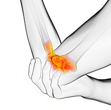 A Lateral Condyle Fracture refers to the second most common children’s elbow injury. Injuries like these involve a higher risk of nonunion, malunion, and AVN than any other fractures in the pediatric elbow. The Humerus refers to the long bone of the upper arm that extends from the elbow to the shoulder. And lateral condyle fracture refers to a fracture in the lower part of this bone, near the elbow.
It is a common injury in younger children. It often occurs because of falling straight onto an outstretched hand or a direct blow to the elbow. Also, this could happen in falls from scooters, monkey bars, skates, etc.
Causes of Lateral Condyle Fractures in Children
A lateral condyle fracture in children involves a break in the outer portion of the distal humerus, near the elbow. The break typically affects the growth plate (physis) and the capitellum. This injury ranks among the most common elbow fractures in children, usually occurring between the ages of 4 and 10. Understanding what causes it plays a crucial role in preventing and treating the condition promptly.
Below are the primary causes of lateral condyle fractures in children, highlighting the mechanisms, contributing factors, and predisposing conditions.
Trauma from Falls
Mechanism: The most frequent cause occurs when a child falls on an outstretched hand (FOOSH). The force travels through the forearm while the hand remains extended and the elbow slightly bent. This motion drives the radius into the capitellum, generating pressure that fractures the lateral condyle.
Common Scenarios: Children often experience such falls while playing on playground equipment, riding bikes or scooters, or running. Their immature bones and open growth plates make the lateral condyle more vulnerable to this type of force.
Example: A 6-year-old falls off a swing and lands on an outstretched arm, which drives force into the elbow and causes a lateral condyle fracture.
Direct Trauma to the Elbow
Mechanism: A direct blow to the outer side of the elbow can fracture the lateral condyle. Though less common than FOOSH injuries, this type of trauma applies compressive force that disrupts the bone and growth plate.
Common Scenarios: Such trauma often happens during contact sports, accidental collisions, or incidents involving impact with a heavy object.
Example: A 7-year-old playing soccer collides with another child and takes a direct hit to the elbow, resulting in a lateral condyle fracture from the compressive force.
QUESTIONS AND ANSWERS
What is a lateral condyle fracture, and how does it happen in children?
- Lateral Condyle Fracture: A lateral condyle fracture refers to a break in the bony prominence on the outer side of the lower end of the humerus bone (the upper arm bone) where it forms the elbow joint. Orthopedic doctors consider this fracture one of the most common in children.
- Mechanism: Lateral condyle fractures often result from a fall onto an outstretched hand with the elbow slightly bent. The force from the fall transmits to the lateral condyle, leading to a fracture. These fractures occur frequently in children because their bones continue to develop and appear more susceptible to this type of injury.
How is a lateral condyle fracture in a child diagnosed and treated?
- Diagnosis: Lateral condyle fractures are typically diagnosed through a physical examination and imaging studies, such as X-rays. The fracture is classified based on its displacement and alignment.
- Treatment: The treatment approach depends on the severity of the fracture:
- Non-Displaced Fractures: Non-displaced fractures may be treated with immobilization in a cast or splint for several weeks, allowing the bone to heal.
- Minimally Displaced Fractures: Minimally displaced fractures may require closed reduction (realigning the bone without surgery) and casting.
- Severely Displaced Fractures: Severely displaced fractures may require surgical intervention, such as open reduction with internal fixation (ORIF) to reposition and stabilize the fracture with pins or screws.
- Rehabilitation: Physical therapy is an important part of recovery for children with lateral condyle fractures. It helps restore elbow function, strength, and range of motion.
What are the potential complications and long-term outcomes of lateral condyle fractures in children?
- Growth Disturbance: One of the main concerns with lateral condyle fractures pertains to potential growth disturbance in the affected bone. If the fracture involves the growth plate, the patient becomes subject to monitoring for growth problems.
- Stiffness or Reduced Range of Motion: Depending on the fracture and treatment, some children may experience stiffness in the elbow or a reduced range of motion. Early and appropriate rehabilitation can help minimize these issues.
- Nonunion or Malunion: In rare cases, fractures may not heal properly (nonunion) or may heal in a misaligned manner (malunion). Timely and appropriate treatment helps reduce the risk of such complications.
Children with lateral condyle fractures require careful evaluation, treatment, and follow-up to ensure proper healing and minimize the risk of complications. Consulting with a pediatric orthopedic specialist becomes important to determine the best course of action based on the specific fracture’s characteristics and the child’s age and growth stage.
When children break bones, parents need to take them to the very best doctors. At the Medical City Children’s Orthopedics and Spine Specialists, we are the best. We specialize in children and their bones.
Management Of Different Types Of Lateral Condyle Fractures
Type I
The degree of displacement determines the management approach. Doctors manage Type I fractures—those with less than 2 mm of displacement—without surgery by using cast immobilization. They typically apply a long arm cast or splint with the elbow flexed between 60 and 90 degrees. Physicians instruct patients to return within one week for follow-up radiographs taken out of the cast or splint to check for any increase in displacement.
While Marcheix et al reported no progression, Pirker et al observed displacement requiring surgery in 5 out of 51 cases. Doctors usually immobilize the arm for four weeks, but they rely on radiographs to confirm healing before discontinuing the cast. Current literature highlights the need for close follow-up because even minimally displaced lateral condyle fractures can shift. To ensure fracture stability, providers recommend serial radiographs, ideally out of the cast. By detecting early displacement, clinicians can avoid delayed surgery and improve outcomes.
Type II
When displacement increases, but the articular hinge remains intact, doctors typically perform closed reduction followed by pinning. Surgeons place two or three pins either percutaneously or subcutaneously, weighing the pros and cons of each method. Achieving an anatomic articular reduction remains critical for a successful outcome, so surgeons should rarely use open reduction. During surgery, they use arthrography to evaluate the quality of the reduction.
For Type II fractures, the preferred approach remains closed reduction with percutaneous pinning. Surgeons must ensure and maintain joint congruity, often confirmed by intraoperative imaging. When planning treatment, surgeons also consider the patient’s age, fracture characteristics, and potential for growth disturbances. If closed reduction fails or the joint surface remains incongruent, they proceed with open reduction.
Type III
To treat Type III fractures, surgeons perform open reduction and pinning. They typically use a lateral approach and dissect closer to the anterior part of the joint. To avoid avascular necrosis, they take great care to preserve soft tissue on the posterior aspect of the fragment. The reduction must restore full articular congruence. After surgery for Type II or III fractures, doctors apply a long arm cast or splint with the elbow flexed at 90 degrees and the forearm in a neutral position.
They generally remove the cast and pins at four weeks if radiographs show adequate healing, then begin early active range of motion. Most experts agree that open reduction and internal fixation offer the best results for Type III fractures, which usually involve more than 4 mm of displacement and disrupted articular surfaces. To minimize the risk of avascular necrosis, surgeons avoid extensive posterior dissection.
After confirming sufficient healing, they promote early mobilization to restore function and reduce joint stiffness. Radiographic follow-up helps monitor for complications such as nonunion or malunion.
Lateral Condyle Fracture Symptoms
Get immediate medical attention in the following cases:
- Pain is increasing quickly without warning
- Swelling for no reason
- New redness and warmth are being generated around the wrist
- New fevers, chills, nausea, sick feeling
- Pain that does not get better even after taking ibuprofen (Advil®), acetaminophen (Tylenol®), etc.
- Inability to wiggle fingers, numbness
All the above symptoms could appear as signs of a different problem. In that case, you must take your child to the clinic or the emergency department. Keep in mind, certain types of elbow injuries in children, like lateral humeral condyle fractures, appear sneaky. These are the second most common pediatric elbow fractures, especially after a fall onto an outstretched hand. If your child is experiencing swelling, pain, difficulty moving the elbow, or tenderness on the outside of the elbow, don’t ignore it.
Sometimes, you might even notice bruising (ecchymosis) around the outside of the elbow. These injuries are tricky because they can move out of place or fail to heal properly if not treated, potentially leading to lasting problems with elbow movement or appearance. If you notice any of these symptoms, or if your child’s pain is out of proportion to what you’d expect after a minor fall, err on the side of caution and get them checked out right away.
Diagnosis and Treatment of a Lateral Condyle Fracture in a Child
It involves a break in the lateral portion of the distal humerus. It often extends through the growth plate (physis) and into the capitellum. This fracture usually results from a fall on an outstretched hand or direct trauma to the elbow. Due to the involvement of the growth plate and the elbow’s complex anatomy, accurate diagnosis and treatment are crucial to prevent complications like growth disturbance, nonunion, or deformity.
Below is an outline of the diagnostic and treatment processes for a lateral condyle fracture in a child, based on standard pediatric orthopedic practices.
Who Gets Lateral Condyle Fractures?
Lateral condyle fractures most frequently affect children between the ages of 4 and 10, with 6-year-olds being the most common age for this type of injury. Boys appear as a higher risk; about two out of every three cases happen in boys. Injuries are also more likely to involve the left elbow.
Diagnosis of a Lateral Condyle Fracture in a Child
1. Clinical Evaluation:
- History: The child typically presents after a fall, reporting pain in the elbow, difficulty moving the arm, and sometimes a popping sensation at the time of injury. Parents may notice the child avoiding the use of the affected arm.
- Physical Exam:
- Inspection: Swelling or bruising around the lateral aspect of the elbow, with possible visible deformity in displaced fractures.
- Palpation: Tenderness directly over the lateral condyle, often with a grating sensation if the fracture is displaced.
- Range of Motion: Limited elbow flexion, extension, or forearm rotation due to pain. The child may hold the elbow in a flexed position for comfort.
- Neurovascular Assessment: Examination of the radial nerve (assessing wrist and finger extension) and distal pulses ensures no associated nerve or vascular injury.
- Example: A 6-year-old falls off a swing and presents with swelling and tenderness over the outer elbow. In addition, the child cannot fully straighten the arm, prompting further investigation.
2. Imaging Studies:
- X-Rays:
- Standard views include anteroposterior (AP), lateral, and internal oblique radiographs of the elbow. The internal oblique view is particularly useful for visualizing the fracture line. This often runs from the lateral metaphysis through the physis into the joint.
- Fractures are classified using systems like the Milch classification (Type I: fracture exits lateral to the trochlear groove; Type II: exits through the trochlear groove, more unstable) or the Jakob classification (Stage I: <2 mm displacement, non-displaced; Stage II: 2–4 mm displacement; Stage III: fully displaced and rotated).
- Findings: Radiologists can see a displaced fragment, widened physis, or a “fat pad sign” (elevated fat pad due to joint effusion). In non-displaced fractures, doctors need to look very hard as the fracture line requires scrutiny.
- Advanced Imaging:
- MRI: Used for non-displaced fractures to assess cartilage involvement, physeal injury, or soft tissue damage, particularly if X-rays are inconclusive.
What is the clinical value of MRI in evaluating and diagnosing humeral lateral condyle fractures in children?
- MRI for Diagnosis and Evaluation: Magnetic Resonance Imaging (MRI) can play an important role in assessing humeral lateral condyle fractures in children, especially when standard X-rays do not provide a clear picture of the fracture or its extent.
- Advantages of MRI:
- Soft Tissue Assessment: MRI is particularly useful in identifying associated soft tissue injuries, such as cartilage or ligament damage, which may not be visible on X-rays.
- Detection of Occult Fractures: In cases where the fracture is subtle or unclear, MRI can help confirm the diagnosis by revealing fracture lines that are difficult to see with other imaging methods.
- Growth Plate Evaluation: MRI allows better visualization of the growth plate, which is crucial for detecting any involvement that could affect bone development.
- When Is an MRI Used? While X-rays remain the first-line tool for diagnosing most lateral condyle fractures, an MRI can identify fractures. When there is uncertainty. If concern about growth plate damage or additional injuries is suspected, doctors will order an MRI.
MRI is not always necessary, but it adds value in more complex or unclear cases to help guide the appropriate treatment plan for young patients.
- CT: Rarely needed but can help in complex cases to evaluate fragment displacement, joint surface involvement, or surgical planning.
- Arthrogram: Performed intraoperatively to confirm articular surface alignment in displaced fractures, ensuring proper reduction.
- MRI: Used for non-displaced fractures to assess cartilage involvement, physeal injury, or soft tissue damage, particularly if X-rays are inconclusive.
- Example: X-rays of a 7-year-old reveal a Jakob Stage II lateral condyle fracture with 3 mm displacement, best seen on the oblique view, with a positive fat pad sign indicating joint effusion.
3. Differential Diagnosis:
- Doctors need to look for other elbow injuries, such as supracondylar humerus fractures, medial epicondyle fractures, or radial head dislocations, to rule them out. A thorough physical exam and imaging help differentiate these conditions.
Early vs Delayed Treatment for Displaced Lateral Condyle Fractures
When a child’s lateral condyle fracture of the humerus possesses more than 2 mm of displacement, the timing of treatment becomes especially important. Studies comparing early intervention to delayed care show that addressing injuries sooner rather than later can make a significant difference. Early surgical management—typically within a few days of the injury—tends to yield more reliable healing.
Additionally, it promotes better alignment of the bone and reduces the risk of complications like nonunion or malunion.
On the other hand, postponing surgery can increase the likelihood of stiffness and extended healing times. This occasionally necessitates more complex procedures down the road. For this reason, orthopedic surgeons often recommend prompt evaluation and intervention when notable displacement is present. Acting quickly supports the bone’s recovery and improves the chances that your child will regain full movement in the elbow.
How is a Lateral Condyle Fracture Treated
Usually, in these injuries, either the fractured bones are separated/displaced, or are nondisplaced (the bone fragments have not moved). When looking for a second kind of fracture, Doctors and radiologists find it difficult to find them on an X-ray. This is because, in some cases, the fracture line is hardly visible. Surgery is usually necessary when the bone is widely displaced. In surgery, the doctors will move the bone and set it back to its normal position with pins or screws.
Thus, it can heal properly. Your child will wear a cast for protection. To leave room for swelling, the doctor will probably start with a split cast on the arm. In this case, he will tape the sides of the cast with cloth medical tape. These tapes are available at the pharmacy. And you can replace them if the tape starts to peel off. Also, you may use cloth athletic tape or duct tape if your child doesn’t possess a latex allergy. The cast sits firmly in place from the inside. That is why it won’t fall apart even if the tape starts to peel.
Doctors usually do not use waterproof material for these first casts because of swelling. In Particular, swelling in the fingers is quite common. Your child might need help with keeping their arm and hand lifted. The arm should rest above the heart to help with swelling. Finally, treating this type of broken bone takes experience and expertise. Our doctors at Medical City Children’s Orthopedics and Spine Specialists are experts children with Lateral Condyle Fractures. If not us, we strongly suggest you see someone who treats this condition regularly.
The differences between the anterolateral and posterolateral surgical approaches
When surgery is necessary for a lateral condyle child’s fracture, surgeons typically use either the anterolateral or the posterolateral approach. While both techniques aim to properly align the bone so it can heal, there are a few important distinctions:
- Anterolateral Approach: In this method, the surgeon makes an incision toward the front and outer side of the elbow. This provides clear access to the fractured area and allows for careful repositioning of the bone. The anterolateral approach is sometimes preferred because it reduces the risk of damaging important nerves near the elbow joint.
- Posterolateral Approach: This involves an incision at the back and outer part of the elbow. While it can also give good exposure for fixing the fracture, the posterolateral approach might need extra expertise as it may appear slightly closer to certain nerves and blood vessels.
Ultimately, the choice of approach depends on several factors, such as the exact location of the fracture, the child’s anatomy, and the surgeon’s experience. The goal with either method is always the same: to set the bone securely for the best possible healing and to minimize risks.
Closed Reduction Versus Open Reduction: What Does the Latest Research Say?
Recent meta-analyses and clinical studies explored treatment options for pediatric humeral lateral condyle fractures, particularly comparing closed reduction with percutaneous pinning (CRPP) to open reduction with pin fixation (ORIF) for cases with significant displacement (more than 4 mm).
The evidence suggests that, when technically feasible, CRPP provides outcomes similar to those of ORIF in terms of fracture healing and long-term function. Some highlights from the research:
- Comparable Clinical Outcomes: Both CRPP and ORIF show similar rates of successful bone healing and return to normal limb function when performed appropriately for the degree of fracture displacement.
- Reduced Soft Tissue Trauma: CRPP often results in less disruption to the surrounding soft tissue and potentially lowers the risk of complications like infections or growth disturbances.
- Surgical Considerations: In situations where closed reduction is not possible, such as when an anatomic reduction appears unattainable, transitioning to an open approach ensures the best chance for optimal joint congruence and long-term elbow function.
Ultimately, the decision between CRPP and ORIF depends on the specific fracture pattern, the amount of displacement, and the ability to achieve accurate reduction. Consultation with a pediatric orthopedic surgeon skilled in both techniques is important for tailoring care to your child’s needs.
Kirschner Wires vs. AO Cannulated Screws: Which is Better?
When it comes to fixing displaced lateral humeral condyle fractures in children, surgeons may use either Kirschner wires (commonly called K-wires) or AO cannulated screws. Both methods aim to hold the broken bone firmly in place so that it heals correctly, but there are a few differences worth noting.
- K-wires are thin, smooth metal pins inserted through the bone fragments to provide stability while the bone heals. They are often used because the procedure is relatively quick and doesn’t require a large incision. The wires usually need removal after a few weeks, which is a simple procedure but does involve a return visit to the doctor’s office.
- AO cannulated screws are a bit more robust. These screws provide very stable fixation and may allow for slightly faster healing or greater security in holding the bone fragments together. However, placing screws is a bit more involved than placing K-wires, and removing them may require another minor procedure if the screws are not left in place permanently.
Recent studies comparing these two techniques show that both offer excellent outcomes in terms of bone healing and restoring function in the elbow. Ultimately, the choice often depends on the surgeon’s preference and the specific details of the fracture.
Both methods reveal high success rates, so parents can feel confident that their child will heal well with either approach under experienced care.
Surgical Approaches and Outcomes for Lateral Humeral Condyle Fractures
When surgery is recommended for lateral humeral condyle fractures in children, several techniques are available, each with its own benefits. Let’s look at how these approaches compare:
- Open vs. Minimally Invasive Procedures: Some surgeons use a traditional open approach, while others may opt for less invasive options like arthroscopic-assisted reduction. The minimally invasive techniques can lead to smaller scars and potentially quicker recovery times; however, both methods reveal good results in terms of bone healing and restored elbow function.
- Internal Fixation Methods: The way the bone fragments are held together inside the elbow can also differ:
- Kirschner Wires (K-wires): These slim metal pins are commonly used to secure the bone. They’re effective, straightforward to insert, and widely available, but sometimes require removal after healing.
- Biodegradable Pins: These pins dissolve over time, reducing the need for removal surgery later. Studies suggest that healing rates and functional outcomes are similar to K-wires, though individual surgeon experience may influence the choice.
- Cannulated Screws: Some surgeons prefer small screws, like the AO cannulated type, to achieve a snug fit and strong hold, particularly in more displaced fractures. This option may allow children to return to activity sooner, though it is also usually followed by routine monitoring to ensure proper healing.
- Outcomes: Overall, most surgical techniques, when performed by experienced hands, lead to successful healing and good return of elbow motion. Your child’s surgeon will choose the method based on the specific type and position of the fracture, as well as their comfort with each device.
If you have questions about which technique the surgeon will use for your child, don’t hesitate to ask your doctor for their reasons. Your orthopedic team at Medical City Children’s Orthopedics and Spine Specialists stays up-to-date on these procedures to offer the best care possible.
What are the possible complications of untreated or improperly managed lateral humeral condyle fractures?
- Elbow Deformity: If left untreated or improperly managed, lateral condyle fractures can lead to noticeable deformities around the elbow joint in children. This may result in abnormal angulation or shape of the elbow, affecting both appearance and function.
- Nonunion and Malunion: When a fracture doesn’t heal (nonunion) or heals out of alignment (malunion), children may experience persistent pain, swelling, or loss of normal arm function. These issues are more likely if the bone fragments are displaced or not stabilized correctly during healing.
- Restricted Range of Motion: Improper healing can cause the child to lose movement in the elbow, especially if scar tissue develops or the bone heals in the wrong position.
- Avascular Necrosis: In rare cases, the blood supply to the bone fragment may become compromised, increasing the risk of bone tissue death and permanent joint problems.
- Infection (if surgery is needed): Post-surgical complications, such as infections, although rare, can occur, emphasizing the importance of expert care and sterile technique
- Prominent Lateral Condyle: The most frequently reported complication after a lateral condyle fracture is the development of a visibly prominent lateral condyle. This occurs in over a quarter of cases, but fortunately, it generally does not impact the child’s function or long-term clinical outcome.
- Severity and Complication Rates: Children with less severe fractures tend to possess fewer problems down the line. For instance, complication rates are lower in less displaced fractures treated with surgery, while more severe fractures are associated with a higher risk for complications overall.
Striving for a fast and complete recovery
These potential complications highlight why precise evaluation and expert treatment, like that offered at Medical City Children’s Orthopedics and Spine Specialists, are essential in helping young patients achieve the best possible recovery. Early intervention and close follow-up care make a big difference in the outcome.
Children with lateral condyle fractures require careful evaluation, treatment, and follow-up to ensure proper healing and minimize the risk of complications. Consulting with a pediatric orthopedic specialist is important to determine the best course of action based on the specific fracture’s characteristics and the child’s age and growth stage.
When children break bones, parents need to take them to the very best doctors. At the Medical City Children’s Orthopedics and Spine Specialists, we are the best. We specialize in children and their bones.
What are the complication rates associated with different types of pediatric lateral condyle fractures?
Complications tend to occur more frequently with more severe fracture types. For instance, children with less severe (type II) lateral condyle fractures—especially when treated surgically—generally experience lower complication rates, around 11%. In contrast, those with the more severe, completely displaced (type III) fractures may face complications in about a third of cases (close to 34%).
This pattern emphasizes why careful assessment of the fracture type is so important in predicting a child’s recovery and potential risks.
Varus Deformity: Causes and Considerations
Varus deformity, or inward angulation of the elbow, is a known complication that can develop after a lateral condyle fracture in children. This typically arises when the fracture heals in a misaligned position, often due to inadequate reduction or fixation during the initial management. If the lateral segment of the bone does not unite properly, the natural alignment of the elbow may not heal properly, leading to the characteristic “gunstock deformity”.
Factors that increase the risk of varus deformity include:
- Delayed or Insufficient Treatment: If the fracture is not recognized early or if the displacement goes untreated, improper healing is more likely.
- Growth Plate Involvement: Damage to the growth plate can alter normal bone growth, resulting in angular deformities as the child matures.
- Nonunion or Malunion: Failure of the bone ends to join (nonunion) or healing in a poor position (malunion) directly contributes to the abnormal angulation.
Early radiographic follow-up and precise alignment during treatment significantly reduce the likelihood of developing a varus deformity after a lateral condyle fracture.
Can my Child Stay Active with a Lateral Condyle Fracture?
Despite the cast providing some protection, a direct blow to the arm or elbow could make the fracture worse. To stay safe, your child should avoid doing any activities with a risk of falling or taking a direct hit to the arm.
Such activities include:
- Contact sports such as basketball, hockey, and soccer
- Playing on playground structures such as jungle gyms or swing sets
- Ice skating, horseback riding, or skiing
Will my Child experience discomfort or Pain?
In the first few days through the first week, soreness is usually at its worst. Doctors often recommend acetaminophen (Tylenol®) or ibuprofen (Advil®) to treat pain from soreness. However, always discuss allergies your child may possess before taking over-the-counter medication. Experts often prescribe a small amount of pain medicine post-surgery if necessary.
How Long Will My Child Need a Cast for a Lateral Condyle Fracture?
Commonly, the timeline for this injury is four to six weeks in a long arm cast. After that, your child will again go to see the doctor, who will remove the cast and do an X-ray. Also, they will remove pins from the surgery once the bone heals and is stable. However, if they used screws instead, removing them may not become necessary.
Will my Child Need Treatments or Physical Therapy after Casting?
Usually, most patients do not require physical therapy. Your child will gain complete strength and movement within one or two weeks after the casting ends. However, if there is any trouble restoring full motion, doctors might recommend therapy.
How Long Should My Child Stay Away from Sports?
We evaluate each patient and make recommendations depending on how the fracture looks. Also, we assess the potential injury risks of the sport your child loves to play. This kind of injury may keep patients away from playing contact sports for two to three months.
What is the Right Time to Follow Up?
During the first two weeks, experts often see patients once a week for X-ray checks. Every arm muscle can put pressure on the fractured bone, even though it is immobilized in a cast. This tension tends to make the bone set out of the right position. Specialists carefully monitor this and treat it early if it happens.
If the first cast was split and everything looks alright in the first week of the appointment, experts overwrap the cast. This means adding a new layer of casting material to close off the cast. This prevents it from getting too loose as swelling is relieved.
When Should I Contact the Doctors?
Get immediate medical attention in the following cases:
- Pain is increasing quickly without warning
- Swelling for no reason
- New redness and warmth are being generated around the wrist
- New fevers, chills, nausea, sick feeling
- Pain that does not get better even after taking ibuprofen (Advil®), acetaminophen (Tylenol®), etc.
- Inability to wiggle fingers, numbness
All the above symptoms could identify the signs of a different problem. In that case, you must take your child to our clinic or the emergency department. Proper medical attention, a little patience with care, and your child will get better and better.
Why See the Doctors at Medical City Children’s Orthopedics and Spine Specialists
Every year, hundreds of children, teens, and young adults with fractures of all sorts and degrees of complexity are treated by the orthopedics and fracture experts at Medical City Children’s Orthopedic and Spine Specialists with offices in Dallas, Arlington, Flower Mound, Frisco, and McKinney, TX.
Furthermore, our expertise in children allows us to diagnose diseases affecting the growing musculoskeletal system and establish appropriate treatment plans. Orthopedic injuries that require immediate medical attention are treated by our orthopedic specialists. We invite you to give us a call and make an appointment.
We specialize in children and are experts in treating them for all musculoskeletal conditions.
____________________
Citation: Journal of Bone and Joint Surgery – Lateral Condyle Fracture
The medical content on this page has been carefully reviewed and approved for accuracy by the Medical City Children’s Orthopedics and Spine Specialists’ qualified healthcare professionals, including our board-certified physicians and Physician Assistants. Our team ensures that all information reflects the latest evidence-based practices and meets rigorous standards of medical accuracy, with oversight from our expert spine doctors to guarantee reliability for our patients.
Call 214-556-0590 to make an appointment.
Comprehensive services for children from birth through adolescence at five convenient locations: Arlington, Dallas, Flower Mound, Frisco and McKinney.