
RADIUS ULNA FRACTURES
The radius and ulna are the two bones that make up the forearm. The majority of forearm fractures in children involve both broken bones.
At Medical City Children’s Orthopedics and Spine Specialists, our broken arm Doctors and specifically broken forearm doctors are dedicated to diagnosing and treating children and ensuring comprehensive care tailored to each patient’s needs. With advanced techniques and a compassionate approach, our team is here to diagnose, treat and care for children suffering from Radius Ulna Fractures.
If your child needs surgery or casting, our Fracture Care Clinic opens every day and you do not need an appointment. Surgery rooms get scheduled every morning, so your child receives the care and attention they need right away.
Radius Ulna Fractures
The radius and ulna form the two bones of the forearm. Most forearm fractures in children involve both bones. These fractures usually happen in the middle, near the elbow, or close to the wrist. This article focuses on fractures involving the radius and ulna.
Although most fractures affect both bones, isolated radial shaft fractures can still occur. Doctors rarely see a radial shaft fracture without some injury to the ulna. When the radius alone breaks and stays aligned, doctors may use a cast for healing. If the bone shifts or both bones break, doctors may recommend surgery for proper alignment.
Understanding fracture patterns and treatment options helps you choose the best care for your child’s recovery.
What is a Broken Forearm?
 A forearm fracture means one or both bones between the elbow and wrist have broken. The forearm contains two bones: the ulna and the radius.
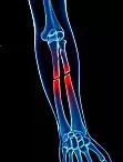
The ulna lies on the pinky side, while the radius sits on the thumb side of the forearm. These bones run parallel, linking the elbow to the wrist.
The ulna is longer and thinner, located on the inner side when your palm faces up.
The radius is thicker and rests on the outer side, next to the thumb. Together, the radius and ulna provide strength and motion to the forearm. They help you twist your arm and perform daily activities like lifting, gripping, or turning.
Kids often fracture their forearms while playing or falling. The forearm is one of the most commonly broken areas in children. Fractures may occur in the bone’s center or near the wrist or elbow. In many cases, both the radius and ulna break together. Fracture location and severity influence treatment plans and recovery time. Your child’s doctor will use imaging and exams to choose the best healing treatment.
QUESTIONS AND ANSWERS
What is the cause of the radius or ulna fracture, and how can we prevent it from happening again?
The cause of a radius or ulna fracture in a child can vary, with common reasons being falls, sports injuries, or other forms of trauma. Accidents are a part of childhood, and it’s challenging to prevent every fracture. However, parents can take some preventive measures, such as ensuring a safe environment, providing proper supervision during activities, and encouraging the use of protective gear during sports or play.
What is the recommended treatment for my child's radius or ulna fracture, and what is the expected recovery time?
The treatment for a radius or ulna fracture depends on the type and severity of the fracture. In many cases, the doctor may recommend immobilization using a cast or splint to allow the bones to heal properly. Simple fractures may not require surgery, but more complex fractures might need surgical intervention. The expected recovery time varies but can range from several weeks to a few months. Rehabilitation, including physical therapy, may also be recommended to restore strength and range of motion.
Are there any potential long-term effects or complications associated with a radius or ulna fracture in children?
Most children with a radius or ulna fracture can expect a full recovery with appropriate treatment and follow-up care. However, potential complications could include issues like malunion (improper healing of the bones), growth plate injuries, or stiffness in the joint. In some cases, more specific complications such as malunion—where the bone heals in an improper position—or nonunion—where the bone fails to heal properly, possibly due to poor blood supply or infection—may occur. Regular follow-up appointments with the doctor are crucial to monitor the healing process and address any concerns promptly. Parents should encourage their children to participate in rehabilitation exercises as recommended to minimize the risk of long-term complications.
It’s important for parents to communicate openly with the healthcare provider, follow their advice regarding treatment and rehabilitation, and attend scheduled follow-up appointments. Each fracture is unique, and the doctor will provide guidance tailored to the specific circumstances of the child’s injury.
The Doctors at Medical City Children’s Orthopedics and Spine Specialists are experts in treating children and adolescents for Radius Ulna Fractures. See the Specialists for children.
Radius Ulna Fracture Symptoms
Symptoms of a broken arm may include:
- Acute, immediate agony
- Swelling and sensitivity
- Forearm, hand, or elbow numbness
- Forearm, elbow, or wrist deformity
- Having trouble rotating or twisting the forearm
Signs of Nerve or Blood Vessel Injury
When a forearm fracture occurs, it’s important to watch for symptoms that could indicate injury to the surrounding nerves or blood vessels. Prompt recognition of these signs helps ensure your child receives the right care and avoids further complications.
Warning signs to monitor include:
- Numbness or tingling in the fingers or hand, which may mean nerve involvement
- Pale, cool, or bluish skin in the hand or fingers, suggesting reduced blood flow
- Difficulty moving the fingers or wrist
- Intense pain, especially when moving the forearm or fingers
- Swelling so severe that it causes a tight or “full” feeling in the forearm
If you notice any of these symptoms following a forearm injury, seek immediate medical attention. Early treatment will prevent long-term problems.
How are Ulnar Shaft Fractures Diagnosed?
To determine if your child has an ulnar shaft fracture, doctors will typically start with a physical exam. Then they will discuss any recent injuries or accidents that may have caused the pain. The gold standard for diagnosis is an X-ray. The care team will take images of the entire forearm, including the wrist and elbow, to get a complete picture of the injury. This helps them not only spot any breaks or cracks in the ulna but also check for related surrounding injuries.
During the evaluation, the doctor will pay close attention to alignment and any displacement of the bone. The doctor will also look for any injury to the growth plates. A clear set of X-ray images from different angles, usually front (AP), side (lateral), and sometimes oblique views, ensures the most accurate assessment. If there’s any uncertainty or the fracture appears complex, your doctor may order additional imaging. For instance, a CT scan is used to get a better look at the injury and plan the best treatment strategy.
How is a Radial Shaft Fracture Diagnosed?
Diagnosing a radial shaft fracture usually starts with reviewing how the injury happened, followed by a physical exam. Your child’s doctor will examine the arm for deformity, tenderness, swelling, or other visible injury signs. To confirm the fracture and assess severity, the doctor will order X-rays of the forearm, elbow, and wrist. Sometimes, the initial X-rays don’t clearly show the fracture. In those cases, your doctor may order additional imaging to get better views. This may include delayed X-rays, an MRI, or a CT scan for clearer bone and tissue images. These advanced tests help detect hidden fractures or related injuries, such as damage to the scaphoid or wrist.
Accurate diagnosis helps guide effective treatment and promotes proper healing. Your child’s specialist will also plan rehabilitation to restore arm strength and full movement. To properly diagnose the fracture, doctors begin with history-taking, physical exam, and X-rays from multiple angles. Standard X-rays usually show the forearm, wrist, and elbow to locate the break. If the break isn’t visible, your doctor may repeat X-rays or order an MRI or CT scan. These scans reveal small fractures and confirm any injury to nearby structures like the scaphoid. This detailed evaluation helps your doctor create the best possible treatment plan for your child.
Diagnosing Both-Bone Forearm Fractures
Doctors rely on X-rays to make a clear diagnosis when both forearm bones may appear broken. Typically, they order standard X-ray images, specifically, anteroposterior (front-to-back) and lateral (side) views of the forearm. These images are taken with the arm positioned straight and the palm facing up, allowing the orthopedic team to see both the radius and ulna.
This careful imaging helps pinpoint the exact location and severity of the breaks, reveals whether any dislocation has occurred, and guides decisions on the best course of treatment. In some cases, additional imaging studies might be recommended if the injury is more complex or involves the growth plate. Prompt and precise diagnosis ensures that both-bone forearm fractures are managed effectively, minimizing long-term complications for growing children and teens.
Different Types of Forearm Fractures
Forearm fractures can affect the radius or ulna, or both. These are referred to as Galeazzi or Monteggia fractures when both bones are broken at separate levels, and there is damage to the joint at the wrist or elbow.
A few special types of forearm fractures are important to recognize:
Galeazzi Fracture
There is a problem with both forearm bones. In the majority of instances, the wrist joint sees an ulna dislocation and a shattered radius. This fracture occurs when the radius breaks independently from the ulna, causing the end of the ulna to become dislocated at the wrist. More specifically, Galeazzi’s fracture happens at the distal radio-ulna junction, where the distal radius is fractured, and the ulnar head dislocates.
Monteggia Fracture
This injury needs rapid medical attention since it affects both forearm bones. Typically, the wrist’s radius is dislocated, and the ulna is broken. It involves a fracture of the proximal one-third of the ulnar shaft along with a dislocation of the radial head at the elbow. Because the elbow is involved, prompt diagnosis and treatment are crucial for proper recovery.
Hume Fracture
A less common but distinct type, the Hume fracture, occurs when there is a fracture of the olecranon (the bony tip of the elbow) accompanied by an anterior dislocation of the radial head. This combination of injuries can complicate elbow movement and requires specialized care.
Plastic Deformation
Usually affects the radius or ulna. Children’s growing bones are more elastic than adults’ bones. With excessive force, a child’s bone can experience deformation (bowing) instead of breaking outright. This deformity then remains after the force is removed.
Nightstick Fracture
These occur when the ulna fractures independently of the radius. Doctors can feel the ulna from the tip of the elbow to the wrist, making it particularly vulnerable when children fall to the ground and land on their elbows.
These varied fractures highlight the complexity of forearm injuries, underscoring the importance of understanding the specific type of fracture for effective treatment and recovery. Each type not only involves different bones and joints but also requires a tailored approach to management, emphasizing why prompt evaluation and specialized care are so vital.
Treatment Options for Nondisplaced or Minimally Displaced Nightstick Fractures
When it comes to nondisplaced or minimally displaced nightstick fractures, the main approach is to immobilize the arm and allow the bone to heal naturally. Typically, doctors use a sugar-tong splint for the first week or so (about 7–10 days) to provide initial support and limit movement. After this period, treatment often shifts to a functional brace paired with gentle exercises to restore motion in the elbow, wrist, and hand.
In certain cases, especially if your child is experiencing minimal discomfort, a simple sling combined with a compression wrap may work well. The goal is always to strike a balance between protecting the bone as it heals and encouraging early, safe movement to reduce stiffness and promote a full recovery. Your orthopedic specialist will tailor the treatment plan to your child’s unique injury and symptoms, ensuring the best possible outcome.
Both Bones and Forearm Fractures for Teens
A patient in their adult years who sustains a fracture to both bones usually needs surgery. Without surgery, the forearm is frequently unstable, making it impossible to cast this kind of fracture. Nonsurgical therapy options are available for younger children, but surgery is normally needed for teens. Placing a metal plate and screws on the radius and ulna bones is the most typical treatment for both forearm bone fractures.
How is a Forearm Fracture Treated?
The manner of treatment for a forearm fracture is determined by the patient’s age, the kind of fracture, and how severe the fracture is. One fundamental principle governs the treatment of shattered bones: the fragmented bones must become repositioned and restrained from shifting as they recover. Proper stabilization of the radius and ulna is important because they support each other. Future issues with wrist and elbow mobility may occur from incorrect bone alignment throughout the healing process.
Ultimately, the primary goals of fracture management are to achieve an acceptable reduction of the bones, provide stable fixation to encourage proper bone healing, ensure an early return to daily activities, preserve function, and minimize complications along the way. Every treatment decision—whether surgical or nonsurgical—centers on these guiding principles, aiming to restore the forearm’s function while reducing the risk of long-term problems. Surgery is usually necessary for adult forearm fractures to align the bones and stabilize them for proper healing.
Immediate Treatment
Depending on how far out of position the fragments are, the doctor may attempt to temporarily realign the bones while your child is still in the emergency department. The precise word for this procedure, in which the doctor arranges the components, is reduction. This is not a surgical procedure.
Closed reduction involves realigning the bone fragments externally. Before this, an anesthetic—typically given through an IV in the arm—helps manage your child’s pain, ensuring comfort during the process. After the bones are aligned, your doctor will put a splint (similar to a cast) on your child’s forearm and give your child a sling to hold the arm in place. A splint, as opposed to a complete cast, may be adjusted for tightness or looseness and allows for swelling to take place without risk. This approach not only stabilizes the fracture but also permits some flexibility in response to any changes in swelling, ensuring your child’s arm is comfortably supported as it heals.
Controlling a shattered bone’s mobility is crucial. Moving a broken bone may result in further injury to the bone, surrounding blood vessels, surrounding nerves, and other tissues. Your child will also receive pain medicine and an ice pack as additional rapid therapy to aid with swelling reduction.
Potential Complications From Immobilization
While immobilization is essential for the healing of forearm fractures, it comes with its own set of challenges, particularly when it comes to the rest of the arm and even the body as a whole. One common complication is the development of “frozen shoulder” (adhesive capsulitis). When the arm remains in a sling or cast for an extended period, the lack of movement can lead to stiffness, inflammation, and a significant decrease in the shoulder’s normal range of motion. Even though the injury may be in the forearm, the shoulder often pays the price for inactivity.
Another concern is osteoporosis, which is the weakening of bones over time. Bones grow stronger in response to regular stress and movement. When the forearm—and sometimes the entire arm—is immobilized, this natural strengthening process is put on pause. In some individuals, especially those already at risk, the reduced use of the limb can lead to faster bone density loss in both the immobilized arm and elsewhere in the body.
Other complications related to immobilization and healing can include:
- Stiffness in nearby joints, like the wrist and elbow, makes rehabilitation longer and sometimes more difficult.
- Decreased blood flow from inactivity, which might slow the healing process or encourage swelling.
- A small risk of nerve or blood vessel complications due to sustained pressure or improper positioning.
- Delayed union or improper healing (malunion/nonunion) if movement occurs before the fracture is stable.
For all these reasons, your healthcare provider will work with you to carefully balance the need for stability with the importance of gentle movement and rehabilitation exercises as healing progresses.
Nonsurgical Treatment for Radius Ulna Fractures
Doctors can treat a fractured bone with only a cast or brace if there is just one broken bone and it is not misaligned. Your doctor will regularly order X-rays at the clinic and closely examine the healing of the fracture. Your child could need surgery to put the bones back together if the fracture moves.
Surgical Treatment for Radius Ulna Fractures
Surgery is often necessary when both forearm bones are broken, or if an open fracture has occurred where the bones have pierced the skin. Open fractures typically require prompt surgery due to the increased risk of infection. In the emergency room, patients frequently get intravenous (IV) antibiotics as well as a tetanus injection. Doctors will meticulously clean out the cuts from the injury during surgery. Usually, the shattered bones are mended during the same procedure.
To thoroughly clean the affected soft tissues, doctors may use several different procedures in cases with severe open wounds. If the skin around the fracture is intact, your doctor may recommend waiting until the swelling subsides before having surgery. Keeping the arm still above heart level for several days reduces swelling. It also gives the overly stretched skin a chance to recover.
While most forearm fractures involve both the radius and ulna, it’s worth noting that injuries to a single bone, such as an isolated fracture of the radial shaft, are uncommon. In these rare cases, surgery is typically required unless the fracture is non-displaced. Non-operative treatment for isolated radial shaft fractures is seldom used, as past outcomes have shown less favorable results with nonsurgical management.
If a nondisplaced fracture is present, the doctor may recommend initial immobilization via a splint, followed by a long arm cast once swelling reduces. However, open reduction and internal fixation remain the standard of care for most radial shaft fractures, with the surgical approach tailored to the exact location of the break.
This thorough approach ensures the best possible alignment and stabilization of the bones, promoting optimal healing and minimizing the risk of future complications with arm function.
With Plates and Screws, Perform an Internal Reduction
Forearm fractures most frequently require this kind of surgical treatment. The doctor initially realigns (reduces) the broken pieces of bone during this treatment. A combination of metal plates affixed to the outside of the bones and specialized screws holds the bones together.
Internal rod fixation and open reduction
In this treatment, a specifically crafted metal rod is inserted by the surgeon through the marrow space in the middle of the bone.
External fixation
Using plates and screws and making wide incisions may worsen the skin’s damage if the muscles, bone, and skin are already badly compromised. This might cause an infection. Your child could receive treatment with an external fixator in this situation. The procedure involves inserting metal pins into the bone above and below the fracture location. A bar outside the skin holds the pins and screws. In order to allow the bones to mend, this device acts as a stabilizing frame.
Complications from Radius Ulna Fracture Surgery
Any operation, whether it is to treat a forearm fracture or anything else, has the danger of infection. Nerve and blood vessel damage in the area of the forearm is a little possibility. Synostosis, a bridge of bone that forms when the two forearm bones heal together, is another uncommon consequence. This may limit the bones’ ability to rotate and prohibit some mobility. Surgery does not ensure that the fracture will heal, and parents should understand that further surgery may occur if the fracture does not heal.
The screws, plates, and rods used to repair the fracture may move or shatter — causing the fracture to tear apart. There are several causes for this, including the patient disobeying instructions. Some illnesses, such as diabetes, impede the healing process. Healing is also slowed down by smoking and other tobacco use. Healing is frequently slower if the fracture involves an incision on the skin. Additionally, infections might hinder or delay the healing process.
Depending on the severity and location of the fracture, the elbow or wrist may become stiff. The whole rotation of your child’s palm upward and downward can not function properly. Plates placed over the bone can cause discomfort, especially if placed at the end of the ulna just below the skin. Symptomatic plates may be removed later when the fracture has healed. Loss of reduction is the most typical side effect of non-operative therapy. This indicates that the bone ends closest to the location of the fracture drift apart. Doctors will consider surgery necessary if parents persist with non-operative treatment and healing does not occur.
Potential Complications Following Ulnar Shaft Fractures
While most ulnar shaft fractures heal well with proper care, some complications can arise, especially when the injury is complex or the healing process doesn’t go as planned. Here’s what parents need to keep in mind:
- Nerve and Blood Vessel Injury: There is a risk of damage to nerves or blood vessels near the fracture, although this is uncommon.
- Compartment Syndrome: In rare cases, abnormal pressure can build up within the muscles of the forearm or elbow, restricting blood flow. This condition, known as compartment syndrome, is a surgical emergency due to the potential for permanent damage.
- Delayed or Improper Healing: Sometimes, the broken bone does not heal correctly (malunion) or fails to heal at all (nonunion). Malunion can lead to the bone knitting in an awkward position. When this occurs, it limits function and causes pain due to inadequate blood supply, infection, or fracture site instability.
- Loss of Motion and Stiffness: Immobilization required for healing might cause stiffness in nearby joints, such as the shoulder or wrist. In children, a condition known as “frozen shoulder” is rare but can lead to discomfort and a loss of movement if left untreated.
- Infection: If the bone breaks through the skin or if surgery is required, there is a risk of bone infection (osteomyelitis). This is why keeping wounds clean and following post-surgical care instructions is crucial.
- Bone Weakness: Prolonged immobilization or lack of activity can contribute to the bone becoming weaker (osteoporosis), especially if healing is slow.
- Rare Syndromes: Some children might develop complex regional pain syndrome, which causes chronic pain and changes in skin color and temperature.
Conclusion
The good news: With attentive orthopedic care and following your child’s recommended treatment plan, most complications can be minimized or prevented. If you notice unusual swelling, severe pain, numbness, or if your child’s arm looks very different during healing, contact your doctor promptly.
What Are the Possible Early and Late Complications of Radial Shaft Fractures?
With any orthopedic injury, it’s important to understand the potential complications that can arise after a radial shaft fracture. These complications fall into two main categories—early and late—based on when they tend to appear during recovery.
Early complications:
Before the bone even has a chance to begin healing, there are a few problems that may show up. These include:
- Compartment syndrome is a rare but serious condition where swelling leads to increased pressure within the muscles. This threatens the blood supply and function and requires urgent attention.
- Neurovascular injury: Nerves or blood vessels near the fracture may incur damage during the injury or while the doctor repairs the fracture. This can lead to numbness, weakness, or circulation issues.
- Infection: There is always a risk that bacteria can enter the wound or surgical site. Particularly if the fracture is open or if surgery is necessary.
Late complications:
After the initial stages of healing, other issues may become apparent, sometimes months down the road:
- Nonunion: The fractured bone ends fail to heal together, leaving a persistent gap that may require further intervention.
- Malunion: The bone heals, but in the wrong position or alignment, which can affect motion and function.
- Hardware problems: Plates, screws, or rods used in surgery might loosen, break, or cause irritation, possibly necessitating another procedure.
- Infection (again): Deep or chronic infections can still occur even after surgery, sometimes long after the initial injury.
- Synostosis: Abnormal bone growth causes the radius and ulna to fuse, restricting the normal twisting motion of the forearm.
- Stiffness and pain: Ongoing discomfort and loss of motion, particularly when scar tissue builds up or the joint remains immobile for too long.
Understanding these risks allows families and patients to watch for warning signs and seek prompt care, which can make a significant difference in recovery and long-term function.
How Medical City Children’s Orthopedic and Spine Specialists Care For Broken Forearms
The board-certified pediatric orthopedic surgeons and fellowship-trained physicians at Medical City Children’s Orthopedic and Spine Specialists treat children, adolescents, and young adults who have fractures of all complexities. Our expertise gives room for the accurate diagnosis of problems that relate to the growing musculoskeletal system. We will develop optimal care plans that will ensure that your child’s specific condition is catered to.
Our approach is comprehensive and patient-centered. We provide expert diagnosis, personalized treatment, and ongoing care, ensuring that each step is tailored to your child’s needs. Our specialists employ cutting-edge techniques informed by the latest clinical and scientific research. Our doctors ensure that the care we provide is both advanced and effective. We develop optimal care plans that will ensure that your child’s specific condition is catered to. Our team is continually updating its methods with findings from advanced research, allowing us to offer the most innovative and effective treatment options available.
Finally, we offer personalized treatment and urgent pediatric care services at all of our five locations — Dallas, Arlington, Flower Mound, Frisco, and McKinney, TX.
If you notice any symptoms of a broken forearm in your child, don’t hesitate to contact us to avoid complications. Our pediatric orthopedic specialists are ready to provide expert evaluation and treatment. Early consultation can make all the difference in ensuring a smooth, complete recovery for your child.
____________________
Citation: American Academy of Orthopaedic Surgeons – Forearm Fractures in Children:
The medical content on this page has been carefully reviewed and approved for accuracy by the Medical City Children’s Orthopedics and Spine Specialists’ qualified healthcare professionals, including our board-certified physicians and Physician Assistants. Our team ensures that all information reflects the latest evidence-based practices and meets rigorous standards of medical accuracy, with oversight from our expert spine doctors to guarantee reliability for our patients.
Call 214-556-0590 to make an appointment.
Comprehensive services for children from birth through adolescence at five convenient locations: Dallas, Arlington, Flower Mound, Frisco, and McKinney.