Understanding an Unfused Collarbone in Children
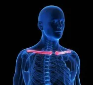 An unfused collarbone, also known as a congenital pseudarthrosis of the clavicle. Additionally, this condition rarely occurs in children, where the clavicle (collarbone) fails to fully form or fuse during fetal development. When this occurs, it results in a gap or discontinuity in the bone. Also, this condition does not appear as a typical fracture, as it presents at birth, but is later found in childhood. For parents navigating this diagnosis, understanding the condition, its implications, and related topics—such as associated conditions, treatment options, and long-term care—is crucial. This article provides a detailed overview of an unfused collarbone. Hopefully, it will provide parents with the information they need as they seek clarity and guidance. Additionally, we focus on why parents should choose Medical City Children’s Orthopedics and Spine Specialists as their first choice for their child’s care.
Causes
The cause of congenital pseudarthrosis remains unclear. Genetic and developmental factors likely contribute. For instance, a family history suggests genetic predisposition. However, scientists and researchers do not identify a specific gene as the cause. Additionally, abnormal fetal clavicle pressure may disrupt ossification. This could stem from positioning or vascular issues. Moreover, the condition can occur with cleidocranial dysplasia. This rare syndrome affects bones and teeth. It causes delayed or absent clavicle fusion. Other skeletal abnormalities may also appear. Meanwhile, environmental factors like maternal health are not well-established. Research continues to explore these influences.
Symptoms
Symptoms depend on defect severity and activity level. Many children do not display symptoms. Often, routine exams or X-rays reveal the condition. Common signs include a clavicle bump or indentation. Additionally, the shoulder may droop slightly. Arm strength can decrease. During activities like swimming, mild discomfort may arise. Rarely, functional limits or cosmetic concerns develop. For example, a widening gap worsens issues. Similarly, an unstable shoulder girdle causes problems. Parents may notice uneven posture. Movement difficulties are also possible.
Diagnosis
Diagnosis begins with a pediatric orthopedic specialist’s evaluation. Initially, the doctor reviews the medical history. Skeletal anomalies or syndromes are noted. Next, a physical exam assesses clavicle mobility. The doctor will check for tenderness or deformity. And test the shoulder’s range of motion. Strength and asymmetry are documented. X-rays confirm the diagnosis. They show a clear clavicle gap with sclerotic ends. This distinguishes it from fractures. In complex cases, CT scans or MRIs are used. These rule out cleidocranial dysplasia. Additionally, blood tests may detect systemic conditions. Thus, the doctor will ensure a thorough diagnosis.
Non-Surgical Treatment
Most children require no immediate intervention. The condition often stays stable and asymptomatic. Therefore, non-surgical care focuses on monitoring. Further, our doctors believe that symptom management counts. For instance, doctors may advise avoiding contact sports. This prevents trauma. Moreover, physical therapy maintains shoulder strength. Gentle exercises support surrounding muscles. The clavicle remains unstressed. Over-the-counter medications relieve pain. Regular X-ray follow-ups track stability. Consequently, many children adapt well. Their bodies use natural compensatory mechanisms.
Surgical Treatment
Our doctors consider surgery as the last choice in treatment. These include functional impairment, persistent pain, or cosmetic concerns. Typically, it occurs in adolescence. The procedure reconstructs the clavicle. In addition, a bone graft, often from the pelvis, is used. Plates or screws stabilize healing. Under general anesthesia, surgery takes 1-2 hours. It aims for bony union. For cleidocranial dysplasia, doctors may need additional procedures. However, surgery appears less common in younger children. Nonetheless, it’s recommended if the defect worsens. Quality of life impacts also prompt surgery.
Care After Surgery
Post-surgical care ensures proper healing. The arm stays in a sling for 4-6 weeks. Doctors manage pain with ibuprofen or acetaminophen. Parents must clean the surgical site daily. Watch for infection signs like redness or fever. Follow-up appointments remove stitches after 10-14 days. X-rays at 6-8 weeks confirm healing. Meanwhile, activity restrictions last 3-6 months. Sports and heavy lifting are avoided. Additionally, shoulder pressure from backpacks is minimized. Thus, recovery occurs effectively.
Rehabilitation
Rehabilitation begins after 6 weeks. A pediatric physical therapist guides the process. Initially, exercises restore shoulder mobility. Pendulum swings and passive stretching are used. At 8-12 weeks, strength training starts. Resistance bands or small weights rebuild muscles. This ensures shoulder girdle stability. Normal activities resume after 4-6 months. Sports may also return, based on healing. Therapist recommendations guide timelines. Consequently, a tailored rehab plan prevents stiffness. It strengthens the shoulder. Complications are minimized.
Associated Conditions and Parental Guidance
Parents should learn about related conditions. For example, cleidocranial dysplasia causes delayed tooth eruption. Wide foreheads or short stature may appear. Orthopedists and dentists provide care. Additionally, osteogenesis imperfecta increases fracture risk. This brittle bone disorder requires attention. Sports injuries are another concern. The condition may lead to shoulder injuries. Proper protective gear reduces risks. Avoiding early sports specialization helps. Furthermore, growth plate concerns are critical. Clavicle growth plates remain active until late adolescence. Surgery must preserve them. Otherwise, growth issues may occur. Bone health is also vital. Calcium, vitamin D, and checkups promote development.
Why Choose Southwest Scoliosis and Spine Institute
The Southwest Scoliosis and Spine Institute excels in North Texas. Our board-certified pediatric orthopedic surgeons specialize in congenital conditions. We provide precise diagnosis. Tailored treatment plans are standard. Advanced imaging is available in our Dallas, Plano, and Frisco locations. Our child-friendly environment reduces stress. Moreover, our multidisciplinary team includes therapists. Specialists address cleidocranial dysplasia and similar conditions. We prioritize long-term health. Personalized rehabilitation and family-centered care are offered. Serving Fort Worth, Arlington, and beyond, we empower parents. Comprehensive support ensures success.
Conclusion
An unfused collarbone is rare but manageable. Careful monitoring is essential. Informed decisions drive success. Also, non-surgical or surgical options aid recovery. Bone health and sports safety improve outcomes. Finally, the Southwest Scoliosis and Spine Institute offers expert care. Our doctors at Medical City Children’s Orthopedics and Spine Specialists are located in five convenient locations: Dallas, Arlington, Flower Mound, Frisco, and McKinney, TX, for families in the Dallas and Fort Worth area. We restore mobility and confidence. Parents gain guidance for optimal outcomes. Contact us to support your child’s journey.
____________________
Citation: Radiopaedia – Congenital Pseudoarthrosis of the Clavicle
Medical City Children’s Orthopedics and Spine Specialists’ qualified healthcare professionals. This includes our board-certified physicians. The medical content on this page has been carefully reviewed. It was approved for accuracy by the doctors and Physician Assistants. Our team ensures that all information reflects the latest evidence-based practices and meets rigorous standards of medical accuracy, with oversight from our expert spine doctors to guarantee the reliability of our information for our patients.
Recent Comments