
INTRA-ARTICULAR FRACTURE
An intra-articular fracture is a fracture that crosses a joint surface. Such fractures also involve cartilage damage. Fractures to joints are more complicated to treat and heal than simple fractures, as multiple bones are involved. Bone fragments inside the damaged joint may impede healing time and efficacy.
At the Medical City Children’s Orthopedics and Spine Specialists medical practice, our expert Intra-Articular Fracture Doctors and surgeons are dedicated to diagnosing and treating children with all types of medical conditions to ensure comprehensive care is tailored to each patient’s needs. We only specialize in children’s health, and we have undergone advanced training to do so. We treat minor and very major medical conditions with a compassionate approach, and our team cares for patients suffering from an Intra-Articular Fracture.
If your child needs surgery or casting, our Fracture Care Clinic opens every day and you do not need an appointment. Surgery rooms get scheduled every morning, so your child receives the care and attention they need right away.
Intra-Articular Fracture
 Intra-articular fractures refer to fractures in which the fracture line penetrates the joint surface, causing some cartilage injury. Also, the fractures might range from displaced to hairline fractures. Usually, intra-articular fractures appear anatomically minimized and secured firmly to permit early joint mobility. Where not possible, some permanent loss of mobility can occur, and the damage may cause the joint to develop degenerative arthritis. Furthermore, other issues that could arise include reflex sympathetic dystrophy, bony protrusion, deformity, numbness, and others.
Causes of Intra-Articular Fractures
High-impact trauma:
- Car accidents, falls from significant heights, or sports injuries can cause bones to break through the joint surface.
Direct blows to the joint:
- A strong, localized force (e.g., a fall directly onto the knee or elbow) can drive part of the bone into the joint space.
Twisting or rotational injuries:
- Sudden twisting of a joint (common in skiing or football) can lead to fractures inside the joint.
Osteoporosis or weakened bones:
- In older adults or those with bone-weakening conditions, even low-impact injuries can result in intra-articular fractures.
QUESTIONS AND ANSWERS
What constitutes an intra-articular fracture in children, and how does it differ from other types of fractures?
Intra-Articular Fracture: An intra-articular fracture refers to a type of fracture where the break occurs within the joint itself or extends to the joint surface. These fractures appear more complex because they affect the articulating surface of the bone, potentially leading to joint instability, cartilage damage, and long-term joint problems.
How do doctors diagnose and treat intra-articular fractures in children?
Treatment: The treatment approach for intra-articular fractures in children varies depending on the specific fracture, the child’s age, and other factors. Treatment options may include closed reduction (non-surgical realignment), open reduction (surgical realignment), and fixation with hardware such as screws or pins. The surgeon’s goal is to restore the joint’s alignment and provide stability while minimizing damage to the joint surface.
In some cases, the severity and location of the fracture, as well as the involvement of tendons, ligaments, muscles, and bones, may necessitate surgical intervention. Minimally invasive closed-reduction surgery might occur through arthroscopically assisted procedures. This technique allows for realignment with smaller incisions, reducing recovery time and minimizing trauma to surrounding tissues.
Alternatively, the surgeon may opt for open-reduction fixation surgery, which involves a larger incision to directly access and repair the fracture. This method becomes essential for more complex or severe fractures to ensure proper alignment and healing.
What is the long-term outlook for children with intra-articular fractures, and how do doctors prevent complications?
- Long-Term Outlook: The long-term outlook for children with intra-articular fractures depends on various factors, including the type and location of the fracture, the quality of treatment, and the child’s age. Intra-articular fractures can potentially lead to complications such as post-traumatic arthritis or joint stiffness. Regular follow-up with a healthcare provider and adherence to rehabilitation and physical therapy will improve the child’s joint function.
- Complication Prevention: While some complications may occur, appropriate and timely treatment and rehabilitation can help minimize the risk of long-term joint problems. Following the healthcare provider’s recommendations for activity restriction and physical therapy can also contribute to a better outcome.
Intra-articular fractures in children require careful evaluation, prompt treatment, and long-term follow-up to ensure the best possible outcome and prevent complications. It’s crucial for parents and caregivers to consult with a pediatric orthopedic specialist or a healthcare provider experienced in pediatric fracture care to develop an individualized treatment plan and provide the child with the best chances for a full recovery.
The doctors at Medical City Children’s Orthopedics and Spine Specialists, are experts in treating intra-articular Injuries. To ensure your child’s bones heal properly, call us for an appointment
Intra-articular Fractures Commonly Occur in the Following Areas:
Shoulder
It might affect the glenoid and articular cartilage, and also the humerus, scapula, acromion, and coracoid processes. Understanding these anatomical components is crucial when addressing various shoulder conditions.
Common Conditions Treated:
- Acromio-Clavicular Strain: This condition involves the ligaments connecting the collarbone to the shoulder blade, often caused by direct impact or overuse.
- Frozen Shoulder: Characterized by stiffness and pain in the shoulder joint, often developing slowly over time.
- Intraarticular Fractures: These fractures occur within the joint space, affecting the movement and function of the shoulder.
- Shoulder Pain Management: Encompasses various treatments aimed at alleviating pain and restoring function, tailored to the specific condition.
By recognizing these conditions, healthcare providers can effectively target treatment strategies, ensuring each component of the shoulder is addressed for optimal recovery. This holistic view is essential for comprehensive shoulder care.
Hip
It could also affect the articular cartilage of the hip and a piece of the pelvis, as well as the head, upper neck, or head of the femur. While there are no specific hip conditions listed for treatment under this category, doctors need to understand the areas impacted. It could also affect the articular cartilage of the hip and a piece of the pelvis, as well as the head, upper neck, or head of the femur.
This information is crucial for understanding the broader scope of potential issues, even in the absence of explicitly listed conditions. Being aware of these areas helps in recognizing symptoms and seeking appropriate medical advice.
Elbow
It can affect the lower end of the humerus, the upper end of the radial and ulnar collateral ligaments, and the cartilage between the bones. The elbow is a complex joint that numerous things can affect. It can affect the lower end of the humerus, the upper end of the radial and ulnar collateral ligaments, and the cartilage between the bones.
Here are some common elbow conditions managed at the facility:
- Bursitis: Inflammation of the bursa, often resulting in pain and swelling.
- Intraarticular Fractures: Fractures that occur within the joint space, impacting movement and stability.
- Tennis Elbow: A condition characterized by pain and tenderness on the outside of the elbow, typically caused by overuse.
By understanding these conditions, patients can better navigate their treatment options and work towards effective recovery.
Wrist
It can affect the lower ends of the radius and ulna, the carpal bones, and the cartilage between the smaller carpal bones. The wrist is a complex joint that can affect the lower ends of the radius and ulna, the carpal bones, and the cartilage between the smaller carpal bones. This intricate structure is susceptible to various conditions that require specialized treatment.
Common Conditions Treated:
- Carpal Tunnel Syndrome: A condition caused by pressure on the median nerve, leading to pain and numbness.
- Dupuytren’s Contracture: A hand deformity that gradually develops over years, affecting the connective tissue under the skin of the palm.
- Fracture of the Middle Phalanx of the Finger: Breaks in the bones of the fingers can severely impact hand function and require precise treatment.
- Ganglion Cysts: Noncancerous lumps that most commonly develop along the tendons or joints of wrists and hands.
- Tendonitis of the Wrist: Inflammation of the tendons around the wrist joint, often due to repetitive motion.
- Trigger Finger: A condition where a finger gets stuck in a bent position and may snap straight.
Understanding these conditions is crucial for effective diagnosis and treatment, ensuring that the complex interplay of bones and tissues in the wrist remains functional and pain-free.
Knee
It can affect the lower edge of the femur, the patella, the upper edge of the tibia and/or fibula, and the articular cartilage and collateral ligaments. It can affect the lower edge of the femur, the patella, the upper edge of the tibia and/or fibula, and the articular cartilage and collateral ligaments.
When discussing knee-related treatments, several specific conditions are typically addressed:
- ACL Repair: A common procedure to fix a torn anterior cruciate ligament, crucial for stabilizing the knee joint.
- Cruciate Ligament Injury: Injuries that affect any of the ligaments inside the knee, impacting its function and stability.
- Fractured Tibia: A break in the shinbone that can significantly impair mobility and require surgical intervention.
- Knock Knees: A condition where the knees angle in and touch each other when the legs are straightened, often necessitating correction for improved alignment.
These conditions highlight the range of knee issues that may need treatment, emphasizing the importance of understanding both the anatomical areas affected and the specific treatments available.
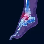
Ankle
It may involve the lower edge of the tibia or fibula, the subtalar joint and articular cartilage, the anterior tibiofibular ligament, and the lateral collateral ligament.
Symptoms of Intra-Articular Fractures
Severe joint pain:
- Pain is often immediate and localized to the injured joint, worsened by movement or weight-bearing.
Swelling and bruising:
- The joint may become significantly swollen and bruised due to internal bleeding and inflammation.
Joint stiffness and loss of motion
- The fracture can disrupt normal joint mechanics, limiting the range of motion.
Visible deformity or joint misalignment:
- In some cases, the joint may appear misshapen or out of position.
Instability or inability to bear weight:
- If the fracture affects load-bearing joints like the knee or ankle, the patient may find standing or walking impossible.
Crepitus or grinding sensation:
- Broken bone fragments may grind against each other during movement.
Diagnosis
Doctors normally diagnose intra-articular fractures through a combination of clinical examination and imaging studies. These imaging tools include visual observation, X-rays, CT scans, and MRI scans. Each of these tests plays a crucial role in the diagnostic process.
- Visual Observation: Initial assessments often involve a thorough physical examination to identify visible signs of injury, such as swelling or deformity.
- X-rays: These are typically the first imaging tests performed, providing a basic view of the bone structure and helping to identify fractures.
- CT Scans: When more detail is needed, CT scans offer a clearer, more detailed image of the bone, allowing doctors to better understand the fracture’s complexity and alignment.
- MRI Scans: These are particularly useful for assessing soft tissue around the joint and any potential impact on ligaments and cartilage.
These tests help assess the location and severity of the fracture, as well as its impact on the joint. By combining these methods, healthcare providers can formulate a comprehensive understanding of the injury, ensuring that the treatment plan is both accurate and effective.
Diarthrodial joints enable the appendicular skeleton’s bones to articulate smoothly and securely, enabling them to perform certain functions. Accordingly, the two end segments of a bone are joined by a fibrous capsule to form a synovial joint. Hyaline cartilage, which is supple, elastic, but avascular, covers the bone’s articulating end portion. Overall, cartilage aids in distributing forces to the subchondral bone underneath. At various joint motion locations, various surfaces of the joint come into contact. Therefore, the joint’s capsule, surrounding muscles and tendons, ligaments, and form all work together to support the joint. Overall, the surfaces of the articular cartilage are lubricated and fed by synovial fluid. Finally, joint health depends on joint movement and repeated physiological loads.
Mechanism of Intra-articular Injury
Articular fractures often result from one of two damage mechanisms:
Indirect Forces
Ligaments provide resistance to the force that causes a bending moment, which changes the eccentric load into a direct axial overload and fractures the joint surface. Henceforth, the most frequent cause of intra-articular fractures is this mechanism. Usually, a partial articular fracture arises from this. Evidently, a split or shearing fracture is often caused by loading one side of the joint, whereas an avulsion fracture or torn ligament is typically caused by pulling on the ligamentous insertions on the opposing side.
Direct Forces
Axial loading forces cause one component to operate as a hammer on the other, impinging on the articular surface or, in more extreme cases, impinging with fractures to the metaphysis or even diaphysis. The fracture pattern depends on the kind of bone, the bone’s location, and the direction and magnitude of the force. Because of the damage that results from this, multi-fragmentary intra-articular fractures can occur. This injury is comparable to crushing. Serious soft tissue injuries are often related.
Evaluation of Intra-articular Fractures
In the event of high-energy injuries, doctors should examine the patient for related musculoskeletal and non-orthopedic injuries. Nevertheless, many systems or multiple bones can get damaged. Basically, Intra-articular fractures can result in joint subluxation or dislocation as well as severe deformities of the limb. To exclude any neurological or vascular damage, our doctors believe a thorough neurovascular evaluation should occur. Examining any accompanying compartment syndrome should occur.
Treatment of Intra-articular Fractures
The treatment approach for intra-articular fractures in children varies depending on the specific fracture, the child’s age, and other factors.
- Initial Step – Immobilization: The first step in the treatment process is immobilizing the joint to stabilize it. This critical measure helps prevent further injury and sets the foundation for subsequent procedures.
- Imaging and Assessment: Following immobilization, imaging is performed to assess the extent of the fracture and any associated injuries. This step is essential for planning the most effective treatment strategy.
- Realignment Options: Treatment options may include closed reduction (non-surgical realignment) and open reduction (surgical realignment). The surgeon’s goal is to restore the joint’s alignment and provide stability while minimizing damage to the joint surface.
- Surgical Intervention: If imaging reveals torn ligaments or shattered bones, surgical intervention becomes necessary. This may involve fixation with hardware such as screws or pins to ensure proper reconstruction of the affected joint.
By understanding each step, from stabilization to potential surgical repair, caregivers can better prepare for the comprehensive care required for their child’s recovery.
Intra-articular Fracture Guiding Principles
The intra-articular fractures may cause stiffness, deformity, discomfort, and posttraumatic arthritis, according to the current operating room philosophy. Certainly, securing an anatomical reduction of the articular surface, reestablishing joint stability and correct axial alignment, and initiating early mobility are critical to prevent deformity and stiffness. Consequently, animal studies show that sustained passive motion followed by anatomical reduction and stable fixation with interfragmentary compression of an intra-articular fracture promotes hyaline cartilage repair. In addition, it is generally recognized that surgeons can repair an articular cartilage defect through the development of fibrous cartilage.
The following identifies the basic principles of treating intra-articular fractures:
- Joint stiffness comes from the immobilization of intra-articular fractures using a plaster cast.
- Increased stiffness occurs as the outcome of immobilizing intra-articular fractures using a plaster cast, open reduction, and internal fixation.
- Closed manipulation and traction influence depressed articular fragments but do not diminish them.
- The instability that comes from their displacement in major articular depressions appears permanent because they do not fill with fibrocartilage.
- To restore joint congruency, doctors should reduce and securely fix articular fragments.
- To stop the repositioning of the decreased articular segments, surgeons must repair the metaphyseal deficiencies beneath them with a structural bone graft or replacement.
- To position the limbs properly and avoid joint loading, the surgeon should displace and decrease the metaphyseal and diaphyseal.
- To avoid joint stiffness and guarantee articular healing and recovery, doctors should allow mobility. This calls for reliable internal fixation.
Non-operative Treatment
Many different fractures might benefit from this therapy. Presently, the conventional course of therapy requires early mobility after immobilization.
Surgery Should Begin Shortly After Intra-articular Damage
- Open fractures
- Irreducible fracture-dislocations
- Linked with neurovascular injuries
- Articular fractures related to compartment syndromes
Severe soft tissue damage normally occurs in intra-articular fractures, and as a result, surgeons will postpone operations until healing occurs. Delays might range from a few days to two or three weeks. If an excessive amount of delay occurs, the surgeon knows that restoring the joint as quickly as possible will ensure a faster and better result. In these situations, it guarantees the reduction of the articular surfaces while exposing as little skin as possible during external fixation. Aligning and fixing articular fragments should only need minor surgery, and lag screws frequently act as implants. When the delay is expected, any metaphyseal fracture or deformity is not addressed at this stage. Both intra-articular and metaphyseal procedures are performed at the same time in situations when the soft tissue conditions appear good, and recovery is possible in 5-7 days.
Mechanism and Procedure for Fixing Intra-articular Fractures
Preserving the blood flow to all of the bone pieces and the soft tissues provides the strategy for the successful repair of articular injuries. Surgeons accomplish this goal by staging intra-articular and metaphyseal procedures, indirect reduction methods, percutaneous lag screw insertion, and, if necessary, the buttressing of these fractures in hybrid frames. It goes without saying that the skin should appear healthy at the time of surgery. The initial surgical procedure for reconstructing intra-articular fractures involves anatomical reduction of the articular surface. The articular cartilage’s depressed areas need raising and repairing.
After the articular fragments are removed, surgeons will use a bone transplant to repair the defect that remains. In these circumstances, autogenous cancellous bone remains the ideal. To prevent axial overload, surgeons must buttress the metaphysis, and the diaphyseal components must undergo repair to initiate early motion. Before closure, the surgeon should repair other intra-articular structures. In cases of compartment syndrome, vascular repairs, open fractures, etc., doctors may use hybrid external fixation instead of the typical use of plates for buttressing. It’s crucial to avoid tensing up the skin. Closing the synovium and capsule will protect the articular cartilage from drying out. It will also cover any exposed tendon or nerve. After five days, some wounds need secondary closure. Surgeons consider using Skin-covering flaps when skin closure does not appear as an option.
Conclusion
A child’s broken joint requires treatment by an orthopedic surgeon who understands the complex workings of the bones, tendons, ligaments, and muscles. Medical City Children’s Orthopedics and Spine Specialists, with offices in Arlington, Dallas, Flower Mound, Frisco, and McKinney, TX, are familiar with the latest surgical techniques for even the most difficult fractures to treat, including complex unhealed fractures. Our orthopedic specialists provide individualized treatment programs that cover every aspect of your care, including diagnosis, treatment, rehabilitation, and aftercare. The type of fracture your child sustained and the location of the injury will determine your course of treatment.
____________________
Citation: Science Direct – Intra-articular Fractures:
The medical content on this page has been carefully reviewed and approved for accuracy by the Medical City Children’s Orthopedics and Spine Specialists’ qualified healthcare professionals, including our board-certified physicians and Physician Assistants. Our team ensures that all information reflects the latest evidence-based practices and meets rigorous standards of medical accuracy, with oversight from our expert spine doctors to guarantee reliability for our patients.
Call 214-556-0590 to make an appointment.
Comprehensive services for children from birth through adolescence at five convenient locations: Arlington, Dallas, Flower Mound, Frisco and McKinney.