
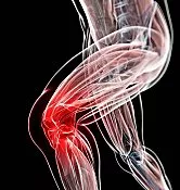
KNEE PAIN
Some of the most common causes of knee pain in kids are dislocations, fractures, sprains, and tears of soft tissues such as tendons and ligaments. Also, in some cases, injuries may involve multiple structures in the knee.
At the Medical City Children’s Orthopedics and Spine Specialists medical practice, our expert Knee Pain Doctors and surgeons are dedicated to diagnosing and treating children with all types of medical conditions to ensure comprehensive care is tailored to each patient’s needs. We only specialize in children’s health, and we have undergone advanced training to do so. We treat minor and very major medical conditions with a compassionate approach, and our team cares for patients suffering from Knee Pain.
If your child needs surgery or casting, our Fracture Care Clinic opens every day and you do not need an appointment. Surgery rooms get scheduled every morning, so your child receives the care and attention they need right away.
Knee Pain
 The common causes of knee pain in kids are traumatic knee injuries, repetitive overuse, and intense physical activities such as competitive sports, etc. Some of the most common injuries that cause knee pain in kids are dislocations, fractures, sprains, and tears of soft tissues such as tendons and ligaments. Also, in some cases, injuries may involve multiple structures in the knee.
It is common for active kids to complain of knee pain even without having sustained any trauma or injury. In most cases, a change in activity level may be the onset of all the symptoms. Knee pain in children and adolescents is a common complaint that can arise from a variety of causes, ranging from benign growth-related issues to serious injuries or medical conditions. Unlike adults, whose knee pain often stems from degenerative conditions like osteoarthritis, children’s knee pain is frequently linked to their developing musculoskeletal system.
Injury Causes, Symptoms, and Diagnosis
Common causes include Osgood-Schlatter disease, a growth-related condition where repetitive stress on the patellar tendon causes inflammation at the tibial tuberosity, often seen in active adolescents during puberty. Patellofemoral pain syndrome, caused by improper tracking of the kneecap, is another frequent culprit, particularly in young athletes involved in sports like soccer or basketball.
Traumatic injuries, such as meniscal tears, ligament sprains (e.g., ACL injuries), or juvenile joint fractures (as you asked about on May 3, 2025), can also cause knee pain, especially in sports-heavy environments. Additionally, conditions like juvenile idiopathic arthritis or infections (e.g., septic arthritis) may present with knee pain, swelling, or stiffness.
Symptoms can range from mild discomfort to severe pain, limping, or inability to bear weight, often requiring prompt evaluation to prevent long-term complications, particularly in growing bones with vulnerable growth plates.
Diagnosis and treatment of knee pain in children and adolescents require a tailored approach, ideally by pediatric orthopedic specialists. Diagnosis typically involves a thorough history, physical exam, and imaging like X-rays to assess fractures or growth plate injuries, or an MRI for soft tissue damage, as seen in conditions like osteogenesis imperfecta or juvenile osteoporosis.
QUESTIONS AND ANSWERS
What are the common causes of knee pain in children?
- Growth-Related Pain: Many children experience knee pain associated with growth spurts, known as growing pains. This is typically benign and often occurs in the evenings or at night.
- Sports-Related Injuries: Active children may experience knee pain due to injuries like sprains, strains, contusions, or fractures from sports or physical activities.
- Overuse Injuries: Repetitive use of the knee joint, as seen in sports or activities like dance, can lead to overuse injuries, such as patellofemoral pain syndrome or Osgood-Schlatter disease.
- Medical Conditions: Conditions like juvenile idiopathic arthritis or Lyme disease can cause knee pain in children. Infections or inflammatory conditions are less common but should be considered.
How can knee pain in children be managed at home?
- Rest: Encourage the child to rest and avoid activities that exacerbate the pain.
- Ice: Apply ice to the affected area for 15-20 minutes every few hours to reduce pain and inflammation.
- Compression and Elevation: In cases of swelling, using a compression bandage and elevating the leg can help reduce discomfort.
- Over-the-Counter Pain Relief:* Non-prescription pain relievers like acetaminophen or ibuprofen, used according to the recommended dosage, can help manage pain. Always consult with a healthcare provider before administering medication to a child.
When should I seek medical attention for a child's knee pain?
- Persistent or severe knee pain that does not improve with rest and at-home care should prompt consultation with a healthcare provider.
- If the child experiences significant swelling, redness, warmth, or an inability to bear weight on the affected leg, immediate medical attention is necessary.
- Knee pain accompanied by other concerning symptoms such as fever, joint swelling, or a rash should be evaluated by a healthcare professional.
- Any suspicion of a traumatic injury or a history of recent falls or accidents involving the knee should be assessed by a healthcare provider.
It’s essential to consider the child’s age, activities, and any underlying medical conditions when addressing knee pain. In cases of persistent or severe pain, or if there is any doubt about the cause, consultation with a pediatrician or pediatric orthopedic specialist is recommended to ensure proper evaluation, diagnosis, and management of the knee pain.
The doctors and surgeons at the Medical City Children’s Orthopedics and Spine Specialists diagnose, treat, and care for children with knee problems that create pain.
We Treat Children with all Types of Knee Injuries, including the Following Major Conditions
Common knee parts that usually get injured are the distal thighbone, proximal tibia, tendons, and ligaments. Generally, most children recover from a knee injury within a few days. Still, it is crucial to know the most common knee injuries and when to see a doctor. Let us take a look:
Knee Fractures
- Knee fractures mean broken or cracked bones in the knee. Also, there might be a dislocated patella or kneecap. A kneecap fracture, also called a patella fracture, is a break in the kneecap, which functions as a shield for the knee joint. Such an injury can be very painful and may impact mobility.
Patellofemoral Syndrome
- Patellofemoral syndrome, often called “runner’s knee,” is a common condition causing pain in the front of the knee. It occurs when the patella (kneecap) doesn’t track properly over the femur (thighbone). This misalignment can result from various factors, including muscle imbalances, overuse, injury, or structural abnormalities. The syndrome causes symptoms like pain during activity, especially when going up or down stairs, squatting, or sitting for extended periods.
Torn Ligaments in the Knee
- Ligament tears occur if a joint is overstretched or twisted. Ligaments can tear totally, or sometimes, it can be a partial tear. It could be an ACL, LCL, PCL, or MCL tear. An incomplete tear feels like a severe strain. But a complete tear can be as painful as a broken bone.
Osgood-Schlatter Disease
- Osgood-Schlatter disease is a common cause of knee pain in adolescents experiencing growth spurts. It affects the tibial tuberosity, the bony bump below the kneecap where the patellar tendon attaches. Repetitive stress and pulling on the tuberosity lead to inflammation and pain. Symptoms are typically exacerbated by activities like running, jumping, and squatting, and the condition usually resolves once the adolescent’s growth is complete.
Juvenile Arthritis
- Juvenile arthritis (JA) is a chronic autoimmune condition that causes inflammation and joint pain in children and adolescents. Unlike adult arthritis, JA is not caused by wear and tear. Instead, the body’s immune system mistakenly attacks its cells, leading to inflammation in the joints. This inflammation can cause pain, swelling, stiffness, and a limited range of motion. JA can also affect other parts of the body, including the eyes, skin, and internal organs. There are several different types of JA, each with its own set of symptoms and potential complications.
Understanding a Torn Meniscus
A torn meniscus refers to an injury involving the cartilage in the knee.
Kneecap Fractures in Children
Kneecap fractures in children are relatively rare but can occur due to trauma. Common causes include:
- Direct trauma to the knee, such as a fall directly onto a hard surface (e.g., playground, pavement).
- Motor vehicle accidents, especially when the knee hits the dashboard.
-
Sports injuries are particularly common in contact sports or activities involving jumping and landing.
- Sudden forceful contraction of the quadriceps, which can pull the kneecap apart, often results in a sleeve fracture (unique to children, involving cartilage).
- Bone disorders, such as osteogenesis imperfecta or conditions affecting bone density, can increase fracture risk.
Symptoms
Signs and symptoms of a kneecap fracture in children include:
-
Severe pain in the front of the knee
-
Swelling and bruising
-
Inability to straighten the leg
-
Difficulty walking or bearing weight
-
Visible deformity or abnormal movement of the kneecap
-
Tenderness over the patella
-
In sleeve fractures, there may be a patellar height change (often looks “high” due to quadriceps pull)
Diagnosis Methods
Physical Examination:
- Assessment of swelling, range of motion, and pain location.
- Palpation to detect movement or displacement of the kneecap.
X-rays:
- Standard imaging to confirm the type and severity of the fracture (transverse, vertical, comminuted, or sleeve).
- Helps determine if the patella is broken into pieces or displaced.
MRI (Magnetic Resonance Imaging):
- It may be used in cases where cartilage or soft tissue injury is suspected (common in sleeve fractures).
Ultrasound:
- Occasionally used in younger children to identify cartilage involvement not visible on X-rays.
Treatment Methods
Non-Surgical Treatment (for nondisplaced or minimally displaced fractures):
Immobilization:
- A knee immobilizer, cast, or splint is used for 4–6 weeks to allow the bone to heal.
Activity restriction:
- The child is advised to avoid putting weight on the leg and to use crutches if needed.
Pain management:
- Acetaminophen or ibuprofen for pain and inflammation control.
Follow-up X-rays:
- To monitor healing progress and confirm bone alignment.
Surgical Treatment (for displaced or complex fractures):
Open Reduction and Internal Fixation (ORIF):
- Re-aligning bone fragments and fixing them with wires, screws, or pins.
Removal of bone fragments:
- In cases where small pieces cannot be reattached.
Rehabilitation:
-
Physical therapy is essential post-surgery or after casting to restore knee strength and range of motion.
🦵 Patellofemoral Syndrome in Children
Runner’s knee, or patellofemoral stress syndrome, results from knee overuse. This injury develops due to repetitive pressure between the thigh bone (femur) and the kneecap (patella). Various factors, such as flat feet, knock knees, muscle imbalances, and improper training, can contribute to this condition. Wearing shoes without adequate support is also a common culprit.
Patellofemoral Syndrome (PFS), also known as “runner’s knee”, is a common cause of anterior (front) knee pain in children and adolescents. It occurs when the patella (kneecap) does not track properly in the femoral groove, leading to irritation of the cartilage or soft tissues surrounding the knee joint.
Causes
Patellofemoral Syndrome is often multifactorial, and common causes in children include:
Overuse or Repetitive Activity:
- Running, jumping, or squatting during sports or physical activities can strain the patellofemoral joint.
Muscle Imbalance or Weakness:
- Weakness in the quadriceps, especially the vastus medialis oblique (VMO), can cause improper kneecap alignment.
Tight Muscles:
- Tightness in the hamstrings, iliotibial (IT) band, or calves can alter knee mechanics.
Poor Alignment or Biomechanics:
- Flat feet, knock knees, or high-arched feet may shift forces in the knee, affecting patellar tracking.
Growth Spurts:
- Rapid growth during adolescence can cause temporary muscle tightness or imbalance, increasing the risk.
Improper Footwear or Training Surfaces:
-
Wearing non-supportive shoes or training on hard surfaces can contribute to abnormal stress on the knees.
Symptoms
Children with PFS often report:
-
Dull, aching pain in the front of the knee
-
Pain that worsens with activity—especially running, stairs, jumping, or prolonged sitting (“theatre sign”)
-
Knee stiffness after rest
-
Popping or grinding sensations (crepitus) during movement
-
Tenderness around the kneecap, especially on the underside
Diagnosis Methods
Medical History:
- Review of symptoms, activity level, and any recent growth spurts or injuries.
Physical Examination:
-
Checking for knee alignment, quadriceps strength, patellar tracking, and muscle flexibility.
-
Special tests like the patellar compression test or Clarke’s sign may be performed.
Imaging (if needed):
-
X-rays: Often normal, but can rule out structural issues.
-
MRI: Rarely needed unless another condition (e.g., cartilage damage) is suspected.
- Note: PFS is usually diagnosed clinically without the need for extensive imaging.
Treatment Methods
Patellofemoral Syndrome is usually non-surgical and responds well to conservative care:
Rest and Activity Modification:
- Avoid activities that worsen symptoms (jumping, deep squats, running).
- Cross-training with swimming or cycling may be recommended.
Physical Therapy:
- Quadriceps strengthening, particularly the VMO.
- Stretching tight hamstrings, calves, and IT band.
- Hip strengthening to improve alignment and reduce knee stress.
- Patellar taping or bracing may help improve tracking temporarily.
Ice and Anti-Inflammatories:
- Ice after the activity to reduce pain and swelling.
- NSAIDs (e.g., ibuprofen) may be used under medical guidance.
Footwear and Orthotics:
- Supportive shoes or custom orthotics for flat feet may be recommended
🦵 Anterior Cruciate Ligament (ACL) Tear in Children
The anterior cruciate ligament (ACL) is one of the four primary ligaments in the knee, crucial for maintaining stability. Positioned diagonally in the knee’s center, it connects the thighbone (femur) to the shinbone (tibia). This ligament is frequently injured, especially in activities involving sudden movements.
Injury to the ACL may happen during pivoting or cutting movements, at the time of landing after jumping, or from a direct blow to the knee. When an ACL is partially or completely injured, the knee might be unstable, or the joint damage can worsen. Such injuries can occur in activities where quick directional changes are required, putting the ACL at risk. If an ACL tear occurs, it can lead to significant knee instability, increasing the likelihood of further joint damage.
Causes
In children, torn ligaments to include ACL tears, are usually the result of non-contact sports injuries, but can also occur due to trauma:
-
Sudden change in direction or stopping quickly (e.g., soccer, basketball, football)
-
Landing awkwardly from a jump
-
Pivoting or twisting with a planted foot
-
Direct impact on the knee (less common)
-
Overuse and improper training in youth sports
In young children, ACL injuries may involve avulsion fractures, where the ligament pulls off a piece of bone instead of tearing in the middle.
Symptoms
Common signs and symptoms of an ACL tear in a child include:
-
A popping sound or sensation at the moment of injury
-
Immediate knee swelling, usually within a few hours
-
Pain and inability to continue the activity
-
Knee instability or giving out, especially with pivoting or sudden movements
- Limited range of motion due to swelling or pain
- Tenderness along the joint line
Diagnosis Methods
Medical History and Physical Exam:
- Review of injury mechanism and symptoms.
- Special tests like the Lachman test, anterior drawer test, and pivot shift test assess ACL stability.
Imaging Studies:
X-ray: Used to rule out bone fractures, particularly avulsion fractures.
MRI (Magnetic Resonance Imaging):
-
Confirms ACL tear.
-
Evaluates associated injuries (e.g., meniscus tear, bone bruises, cartilage damage).
-
Important for assessing open growth plates in children.
Treatment Methods
Treatment depends on the child’s age, activity level, skeletal maturity (open vs. closed growth plates), and severity of the injury.
Non-Surgical Treatment (Selected Cases):
- For younger children with partial tears or those with open growth plates and lower activity demands.
Includes:
-
Physical therapy to restore strength, balance, and motion
-
Bracing to provide knee stability during activity
-
Activity modification to avoid pivoting in sports
-
Close monitoring for any signs of knee instability
Surgical Treatment:
-
Recommended for complete ACL tears in active children, especially those who want to return to sports or have recurrent instability.
Pediatric ACL Reconstruction:
- Uses growth plate-sparing techniques to avoid damaging the physis (growth plate).
- Grafts (e.g., hamstring tendons) are used to reconstruct the ACL.
-
Surgeons choose the technique based on skeletal maturity (e.g., physeal-sparing, transphyseal, or partial transphyseal).
🦵 Medial Collateral Ligament – MCL Injury
An MCL tear usually occurs when a child suddenly twists their knee at the time of running or landing after jumping. It is a sprain or tear of the medial collateral ligament (MCL), a crucial ligament that helps stabilize the inside portion of the knee.
Causes
An MCL tear typically happens during activities involving:
- Sudden changes in direction or twisting the knee while running.
- Awkward landings that twist the knee.
- A direct hit to the outside of the knee while the foot remains planted on the ground.
These injuries are most common in sports that require twisting, cutting, and pivoting, such as skiing, soccer, football, basketball, and tennis. By understanding these causes and symptoms, one can better anticipate and manage the risks associated with MCL injuries.
Symptoms
A popping sound during the injury, followed by knee pain, swelling, and knee weakness, etc.
Diagnosis
To determine if an MCL tear has occurred, a physical examination is essential. Doctors often press on the knee and leg, moving them in specific ways to evaluate ligament integrity. In some cases, imaging tests like X-rays or MRIs may be used to assess the extent of the tear and check for additional damage.
Treatment Options
A variety of treatment strategies can be employed to manage an MCL tear:
- Icing the Knee: Applying ice can reduce pain and swelling. Consider using an ice massage by freezing water in a plastic foam cup, then peeling away an inch of the cup to apply ice directly to the injury for five to ten minutes every 60 to 90 minutes as needed.
- Anti-inflammatory Medications: Over-the-counter medications like ibuprofen or naproxen can help alleviate pain and inflammation.
- PRICE Method: This method involves protecting the knee from further injury, resting, icing, compressing the knee with a bandage or brace, and elevating it above heart level to minimize swelling.
- Adequate Support: Wearing a brace may protect the MCL and knee joint during healing.
- Using Crutches: If necessary, crutches can help avoid placing excessive pressure on the injured knee during recovery.
- Sports Physical Therapy: A physical therapist can address factors like weakness, flexibility, and core strength, guiding your child safely back to activities and reducing future injury risk.
- Surgery: In severe cases, surgical intervention may be required to repair the MCL tear.
These approaches aim to ensure a safe and effective recovery, helping your child regain full function and return to their favorite activities.
🦵 Posterior Cruciate Ligament (PCL) Tear in Children
The posterior cruciate ligament (PCL) is one of the four major ligaments in the knee and is located at the back of the joint. It connects the femur (thighbone) to the tibia (shinbone) and helps prevent the tibia from moving too far backward. Though less common than ACL injuries, PCL tears can occur in children, especially from trauma.
Causes
PCL injuries in children are usually the result of direct trauma or forceful impact. Common causes include:
Direct blow to the front of the knee:
-
Often occurs in car accidents when the shin hits the dashboard (dashboard injury)
-
Falls where the knee strikes the ground while flexed
Hyperextension of the knee:
-
Over-straightening the knee joint can overstretch and tear the PCL
Sports-related trauma:
-
Contact sports such as football, soccer, or rugby
Twisting injury or misstep:
-
It can occur during awkward landings or rapid direction changes
Avulsion injury:
-
In younger children, the ligament may pull off a piece of bone at the tibial attachment (more common than midsubstance ligament tears)
Symptoms
Symptoms of a PCL tear in a child may be subtle compared to ACL injuries and can include:
-
Pain behind the knee after injury
-
Swelling, usually mild to moderate
-
Difficulty walking or bearing weight
-
Feeling of knee instability or looseness, especially when walking downhill or descending stairs
-
Reduced range of motion or stiffness
-
Bruising around the knee in some cases
Mild PCL injuries may go unnoticed at first, as some children adapt to the looseness.
Diagnosis Methods
Medical History & Physical Exam:
-
The doctor will ask about the injury mechanism and examine the knee for swelling, pain, and movement.
-
The posterior drawer test, sag sign, and quadriceps active test are used to assess PCL integrity.
X-rays:
-
Check for avulsion fractures, where a piece of bone is pulled off with the ligament.
-
Useful to rule out other bony injuries.
MRI (Magnetic Resonance Imaging):
-
Gold standard for confirming PCL tears.
-
Helps assess the extent of damage, including involvement of other ligaments or cartilage.
Stress X-rays (in rare cases):
-
It may be used to evaluate posterior tibial translation compared to the uninjured knee.
Treatment Methods
Non-Surgical Treatment
Most isolated, partial, or low-grade PCL tears in children can be managed non-surgically.
Includes:
-
Rest, Ice, Compression, Elevation (RICE)
-
Knee brace (hinged or PCL-specific) to restrict backward tibial motion
-
Crutches to limit weight-bearing initially
- Gradual return to sports with clearance from a healthcare provider
Physical therapy:
-
Strengthen the quadriceps and hamstrings.
-
Improve knee stability and range of motion
Surgical Treatment
Surgery may be recommended if:
-
The tear is complete and symptomatic.
-
There’s a PCL avulsion fracture requiring fixation.
-
Multiple ligaments are torn.
-
The knee remains unstable after conservative treatment
PCL Reconstruction:
-
Involves replacing the torn ligament with a graft (often hamstring or patellar tendon)
-
Pediatric orthopedic surgeons will choose techniques that protect the growth plates in children
🦵 Lateral Collateral Ligament (LCL) Tear in Children
The LCL is a ligament located on the outer side of the knee, connecting the femur (thighbone) to the fibula (outer shinbone). It plays a crucial role in stabilizing the knee during side-to-side movement. While LCL injuries are less common than ACL or MCL injuries, they can still occur in active children, especially those involved in contact or high-impact sports.
Causes
In children, LCL tears are typically caused by trauma that pushes the knee inward while the lower leg moves outward. Common causes include:
-
Direct blow to the inside of the knee (often during contact sports like football, soccer, or hockey)
-
Sudden twisting or pivoting movements
-
Overstretching or hyperextension of the knee
-
Falling awkwardly on a bent knee
-
Severe knee dislocation or multi-ligament injury
In skeletally immature children, the ligament may pull a small piece of bone off the fibula (avulsion fracture) instead of tearing the mid-ligament.
Symptoms
Symptoms of a LCL ligament tear in a child can vary based on the severity and type of the injury, but commonly include:
-
Pain on the outer side of the knee
-
Tenderness along the lateral (outer) joint line
-
Swelling, especially along the outside of the knee
-
Instability or feeling that the knee may buckle during activity
-
Bruising and stiffness
-
Difficulty walking or bearing weight
-
Popping or snapping sound at the time of injury (in some cases)
Diagnosis Methods
Medical History and Physical Exam:
-
The doctor evaluates the mechanism of injury and checks for tenderness, swelling, and instability.
- Varus stress test: Performed to assess laxity in the outer knee when a sideways force is applied.
Imaging Studies:
-
X-ray: Used to check for avulsion fractures or other bone injuries.
-
MRI (Magnetic Resonance Imaging):
-
The gold standard for visualizing the LCL and determining the extent of the injury.
-
Helps identify associated injuries to other ligaments or cartilage.
-
Ultrasound (occasionally used):
-
It may be used in some settings to assess soft tissue injuries dynamically.
Treatment Methods
Non-Surgical Treatment
Most grade I and II LCL tears (mild to moderate) in children can be treated conservatively.
Treatment includes:
-
Rest and avoid weight-bearing activities.
-
Ice packs to reduce pain and swelling
-
Compression and elevation
-
Knee bracing to stabilize the joint.
-
Crutches to assist with walking during early recovery
-
Physical therapy:
-
To restore strength, balance, and range of motion
-
Focus on strengthening surrounding muscles (quadriceps and hamstrings)
-
Children often recover from mild LCL injuries in 4 to 8 weeks with proper rehabilitation.
Surgical Treatment
Surgery is rarely needed, but may be recommended if:
-
The LCL is completely torn (Grade III)
-
There is a bony avulsion that requires fixation.
-
The injury involves multiple ligaments.
-
Chronic instability develops despite conservative treatment.
LCL reconstruction or repair is usually performed using grafts or sutures, especially in high-grade injuries. Pediatric orthopedic surgeons take care to protect growth plates during surgical planning.
Osgood-Schlatter Disease
Osgood-Schlatter Disease is a common cause of knee pain in growing children and adolescents, particularly those who are active in sports that involve running, jumping, or sudden changes in direction. It occurs due to inflammation of the patellar tendon at its attachment point on the tibial tubercle—the bony prominence just below the kneecap. This condition typically arises during periods of rapid growth, when bones, muscles, and tendons are developing at different rates, placing stress on the growth plate. Children with Osgood-Schlatter Disease often experience pain, swelling, and tenderness below the knee, especially during physical activity, but the condition usually resolves on its own with rest, ice, stretching, and activity modification.
Causes Behind Osgood-Schlatter Disease
Osgood-Schlatter disease is primarily an overuse injury targeting the cartilage growth plate located just below the knee, specifically at the tibial tuberosity of the shinbone. This location is crucial as it’s where the patellar tendon attaches to the bone, often under strain from physical activities. The disease can manifest due to ongoing stress on the growth plate or result from a specific incident, such as a fall or a sudden leap. By understanding these triggers, preventative measures can be more effectively implemented.
This condition predominantly arises from repetitive and strenuous movements during physical activities, placing excess stress on the shinbone’s growth plate. Several factors can increase the likelihood of developing Osgood-Schlatter, including:
- Rapid Growth Spurts: During these periods, bones can become vulnerable due to changing length and density.
- Lack of Flexibility: Tight muscles around the knee can lead to increased pressure on the growth plate.
- Sports Activities: Engaging in multiple sports simultaneously or jumping into a new season can exacerbate the risk.
- Continuous Play: Year-round participation in sports without adequate rest can contribute significantly.
Diagnosis
Osgood-Schlatter disease is typically identified through a detailed physical examination of the knee. Healthcare providers assess symptoms and may perform additional diagnostic tests, such as X-rays, to eliminate other possible causes and to check for changes at the tibial tubercle, located just below the kneecap.
Treatment Options
Managing Osgood-Schlatter involves a multipronged approach aimed at reducing pain and allowing the knee to heal effectively. Treatments include:
- Exercise Modification: Temporarily avoid intensive running and jumping to minimize stress on the knee. Focus on low-impact activities like cycling or swimming until symptoms improve.
- Cold Therapy: Applying ice can significantly reduce pain and swelling. Consider using an ice cup massage, moving the ice in circular motions on the affected area for five to 10 minutes, as needed.
- Pain Relief: Use over-the-counter anti-inflammatory medications such as ibuprofen or naproxen to ease discomfort and inflammation.
- Knee Protection: Utilize protective gear such as braces or knee sleeves to cushion and support the knee during daily activities, thereby preventing further strain.
- Physical Therapy: Consulting a sports physical therapist may help correct any underlying physical issues, such as muscle imbalances or tightness, and can assist in gradually returning your child to sports and active play.
This comprehensive plan aims to alleviate symptoms while promoting a safe and effective recovery.
🧒 Juvenile Arthritis (Juvenile Idiopathic Arthritis – JIA)
Juvenile Arthritis (JA), often referred to as Juvenile Idiopathic Arthritis (JIA), is a group of autoimmune and inflammatory conditions that affect children under the age of 16. It causes persistent joint inflammation and stiffness and can impact a child’s overall growth and development.
Causes
The exact cause of juvenile arthritis is unknown. However, several factors are thought to contribute:
Autoimmune response:
-
The immune system mistakenly attacks the body’s joint tissues, causing inflammation.
Genetic predisposition:
-
A family history of autoimmune diseases may increase the risk.
Environmental triggers:
-
Viral or bacterial infections may trigger the disease in genetically susceptible children.
Immune system dysregulation:
-
Improper immune system functioning may lead to chronic joint inflammation.
Note: Juvenile arthritis is not contagious and is not caused by injuries or physical activity.
Symptoms
Many symptoms can vary depending on the type and severity, but commonly include:
-
Joint pain and tenderness (especially in the morning)
-
Swelling and stiffness in one or more joints
-
Reduced range of motion
-
Warmth or redness over affected joints
-
Fatigue
-
Intermittent fevers
-
Limping (especially after waking)
-
Eye inflammation (uveitis) in some forms
-
Poor growth or uneven limb length (from chronic joint inflammation)
Symptoms may come and go in flares and can affect joints symmetrically or asymmetrically.
Diagnosis Methods
There is no single test to diagnose juvenile arthritis. Diagnosis is based on clinical evaluation and ruling out other conditions.
Medical History and Physical Exam:
-
Review of symptoms, onset, duration, and any patterns
-
Assessment of joint swelling, tenderness, and range of motion
Blood Tests:
-
Erythrocyte Sedimentation Rate (ESR) or C-Reactive Protein (CRP): Detect inflammation
-
Rheumatoid Factor (RF) and Anti-CCP antibodies: May be present in some types
-
Antinuclear Antibody (ANA): Often positive in children with eye involvement
-
Complete blood count (CBC): Checks for anemia or elevated white cells
Imaging:
-
X-rays or MRI: To detect joint damage or inflammation
-
Ultrasound: Can reveal joint effusions and soft tissue changes
Eye Exam:
-
Slit-lamp examination: Detects uveitis (a silent but serious complication)
Diagnosis requires symptoms to persist for at least 6 weeks in a child under age 16.
Treatment Methods
The goals of treatment are to control inflammation, relieve symptoms, prevent joint damage, and help the child maintain normal growth and function.
Medications
Nonsteroidal Anti-Inflammatory Drugs (NSAIDs):
-
Ibuprofen, naproxen – for pain and inflammation
Disease-Modifying Antirheumatic Drugs (DMARDs):
-
Methotrexate is commonly used to slow disease progression
Biologic Agents:
-
Target specific parts of the immune system (e.g., etanercept, adalimumab, tocilizumab)
-
Often used if DMARDs are not effective
Corticosteroids:
-
Oral or injected to control severe inflammation or during flares
-
Used sparingly due to side effects in children
Physical and Occupational Therapy
-
Maintains joint flexibility and muscle strength
-
Prevents deformities
-
Customized exercise plans
Regular Eye Exams
-
Especially important for children with positive ANA due to uveitis risk
Surgical Intervention (Rare)
-
Considered in severe cases with joint damage unresponsive to other treatments
Understanding a Torn Meniscus
A torn meniscus refers to an injury involving the cartilage in the knee, known for its important role as a shock absorber and stabilizer. This crescent-shaped cartilage is positioned between the shinbone, or tibia, and the thighbone, also known as the femur. Its primary function is to act as a cushion, providing joint protection and stability during movement.
Common Causes
A torn meniscus can occur due to various reasons, particularly involving movements or impacts that affect the knee joint. In young children, these kinds of injuries are less common but can happen if there is considerable stress or impact on the knee, such as twisting, bending deeply, or experiencing some form of trauma.
- Twisting Motion: Sudden twists or turns on a bent knee can place stress on the meniscus, leading to tears.
- Significant Trauma: Direct force to the knee, whether from sports, falls, or accidents, can result in a meniscus tear.
Uncommon Factors
- Abnormal Cartilage: Some children may have a condition known as a discoid meniscus, which means their meniscus is not the typical crescent shape. Instead, the cartilage forms a more rounded structure, which is prone to snapping or tearing, sometimes without significant force.
Age Considerations
- For children below 12, meniscus tears are rare unless there’s significant trauma or structural abnormalities. When tears do occur, they typically affect only one knee.
Overall, while a torn meniscus in children isn’t as frequent as in adults or teens, knowing the potential causes can help in prevention and identifying symptoms early.
What Are the Symptoms of a Torn Meniscus?
A torn meniscus can significantly affect your daily activities and overall comfort. Here are the primary symptoms to watch for:
- Knee Pain: This is often localized to the area of the tear and can be sharp or aching.
- Reduced Range of Motion: You may find it challenging to fully straighten or bend your knee.
- Swelling: The knee joint might appear swollen or puffed up, sometimes significantly so.
- Locking Sensation: Your knee may feel like it’s catching or getting stuck, leading to difficulty in moving it fluidly.
In addition to these symptoms, you might also experience a popping sensation during the initial injury. If any of these signs persist or worsen, consider consulting a healthcare professional for a thorough evaluation. Early intervention can help prevent further damage and facilitate recovery.
Diagnosing a Torn Meniscus
The first step in diagnosing a torn meniscus begins with a thorough physical examination by a healthcare professional. During this exam, the doctor assesses the knee for swelling, tenderness along the joint line, and range of motion. Special tests, like McMurray’s test, may be conducted to identify meniscal tears by manipulating the knee joint to see if it causes pain or a clicking sound.
Imaging Techniques
To confirm the diagnosis, advanced imaging techniques are often employed:
- Magnetic Resonance Imaging (MRI): This is the most common method used to visualize soft tissue injuries, offering detailed images of the knee’s internal structures. An MRI can provide a clear view of the meniscus and help pinpoint the location and extent of the tear.
- X-rays: Although X-rays do not show meniscal tears, they are useful for ruling out other potential causes of knee pain, such as fractures or arthritis.
- CT Scans: In certain cases, a CT scan may be utilized for a more comprehensive view of the knee, especially if there are complex injuries involved.
By combining physical assessments with these imaging tools, healthcare providers can accurately diagnose a torn meniscus and work with patients on an effective treatment plan.
Knee Sprains
Sprains mean damage to the ligaments that make up the knee. Most sprains result from damage Anterior Cruciate Ligament (ACL) or the Medial Collateral Ligament (MCL). Even any one of these ligaments can lead to severe injuries.
Signs and Symptoms
- Swelling and pain around the knee
- Not able to stand or put pressure on the injured leg
Knee Strains
Just like sprains, strains are common knee injuries in children. However, in this case, the reason is a damaged muscle or tendon within the knee.
Signs and Symptoms
Bruising around the knee and other symptoms similar to a sprain
Tips for Returning to Sports After an ACL Injury
Recovering from an ACL injury and eager to get back on the field? It’s crucial to ensure a safe return by following these essential tips:
1. Prioritize Rehabilitation
-
- Physical Therapy: Engage in a structured physical therapy program. Consistent sessions with a trained therapist can help regain strength and flexibility.
- Home Exercises: Complement your therapy with at-home exercises. Focus on building muscle strength around your knee, which is vital for stability.
2. Embrace Functional Testing
-
- Assess Movement: Functional testing evaluates how well your knee moves and how strong it is, providing insights into your readiness to return to sports.
- Simulate Game Scenarios: These tests often simulate sports-specific activities, allowing for practical assessments of your capabilities.
3. Gradual Return to Activities
-
- Set Realistic Goals: Establish a timeline with achievable milestones. Start with non-contact drills before progressing to full-contact practices.
- Monitor Pain Levels: Keep a close watch on pain and swelling. If discomfort persists, consult your healthcare provider.
4. Invest in Proper Gear
-
- Supportive Braces: Use knee braces if recommended by your doctor to offer additional support during high-impact activities.
- Quality Footwear: Opt for shoes that provide proper cushioning and support to reduce joint stress.
5. Mental Preparation
-
- Build Confidence: Overcome mental barriers by visualizing successful participation in your sport and considering psychological support if needed.
- Patience is Key: Understand that the journey back to full fitness takes time, and rushing the process could lead to setbacks.
Returning to sports after an ACL injury demands dedication and caution. By prioritizing rehabilitation, embracing functional testing, and making informed, gradual progress, you pave the way for a successful and safe return to your favorite activities.
How We Diagnose Knee Pain and Its Associated Knee Injuries
To accurately diagnose a knee injury, our doctors ask about how it happened and assess the associated symptoms. Our child orthopedic specialists will conduct a thorough physical exam by pressing on the knee and legs, moving them in specific ways to reveal the injured part of the knee. To confirm the diagnosis and rule out other possible problems, we recommend imaging tests such as:
- X-rays to check for bone injuries
- CT scans or MRIs to look inside the knee and assess the extent of the damage
Knee Pain Treatment
Treatment for a knee injury depends on its cause and severity. Some knee injuries only require RICE (rest, ice, compression with an elastic bandage, and elevation). However, other knee injuries may need physical therapy, bracing, or even surgery.
For runner’s knee specifically, most cases benefit from:
- Modifying workouts: Avoid activities that exacerbate knee pain. Runners may need to reduce their running distance or change their routes to avoid hills or hard surfaces.
- Icing the knee: This can help alleviate pain and swelling. An effective method is an ice cup massage. Fill a foam cup with water and freeze it. Once frozen, peel an inch of the foam from the bottom and apply an ice massage directly to the injured area for five to ten minutes. Repeat every 60 to 90 minutes as needed.
- Anti-inflammatory medications: Over-the-counter options like ibuprofen or naproxen can help relieve pain and inflammation.
- Adequate support: Ensuring shoes are not overly worn and provide proper support is crucial. A brace, strap, or sleeve might offer relief by stabilizing the kneecap.
- Sports physical therapy: This can be highly effective in eliminating pain and preventing further injury. Strengthening and stretching the quadriceps and hamstrings are key components of rehabilitation.
By combining a detailed diagnostic approach with comprehensive treatment options, we aim to alleviate pain and restore mobility effectively.
Surgical Options Depending on Severity:
The least invasive procedure is arthroscopy, involving small incisions and special tools to repair the knee. Also, there is a knee osteotomy, which fixes one section of the knee. The other surgical options are:
- Plica Removal
- Lateral Release
- Microfracture
- ACL Reconstruction
- Meniscectomy
- Meniscus Repair
- Meniscus Transplant
- Arthrotomy
- Tendon Repair
- Knee Replacements, etc.
By combining thorough diagnostics and a tailored treatment approach, we aim to provide comprehensive care for all types of knee injuries, ensuring effective recovery and a return to normal activities.
Understanding Kneecap Instability (Patella Dislocation)
Kneecap instability, also known as patella dislocation, occurs when the kneecap (patella) slips out of its designated groove at the end of the thighbone (femur). The kneecap plays a crucial role in safeguarding the knee joint and assisting in knee extension. When it shifts from its groove, it results in instability, discomfort, and impaired knee function.
Causes of Kneecap Instability
Several factors can lead to patella dislocation. Typically, it happens when the knee is extended while experiencing a twisting motion. This combination creates enough force to push the kneecap out of position. Additionally, direct impacts to the knee, such as those experienced during contact sports, can also be a common culprit.
Factors that might increase the risk of this condition include:
- Anatomical Variations: Some individuals have naturally shallow or uneven grooves, which can predispose the kneecap to dislocation.
- Muscle Weakness or Imbalance: Weakness in the muscles surrounding the knee can fail to stabilize the kneecap effectively.
- Previous Dislocations: Having experienced a dislocation increases the likelihood of future occurrences.
By understanding these causes, individuals can better gauge their risk and take proactive measures to maintain knee health.
Diagnosing Kneecap Instability
When it comes to diagnosing kneecap instability, the process typically starts with a thorough physical examination. Your child’s physician will evaluate the knee’s range of motion, check for stability, and identify any tender areas.
To gain further insights, X-rays are often used to examine the kneecap’s alignment and detect any fractures involving the kneecap or femur. For a more detailed view, additional imaging such as an MRI or CT scan may be necessary. These advanced techniques can reveal injuries to bones or cartilage, tears in ligaments or soft tissues, and provide a comprehensive assessment of limb alignment.
Treating Kneecap Instability
Immediate Response
After a kneecap dislocation, the patella might realign itself, but in some cases, medical intervention is required.
Non-Surgical Treatments
- Activity Restriction and Immobilization: Limiting movement can prevent further injury and aid in healing.
- Ice Therapy: Applying ice, like using an ice cup massage, can reduce pain and swelling. Freeze water in a plastic foam cup, peel back part of the foam, and massage the area for 5 to 10 minutes. This can be repeated every 60–90 minutes as required.
- Anti-Inflammatory Medications: Over-the-counter medications, such as ibuprofen or naproxen, can help alleviate pain and reduce inflammation.
- Supportive Devices: Braces, straps, or sleeves may be recommended to provide stability and relieve stress on the knee.
- Sports Physical Therapy: A therapist can pinpoint and correct contributing factors like weak quadriceps, poor flexibility, and core strength. They can also guide your child safely back to normal activities.
Surgical Intervention
If cartilage damage within the knee joint results in loose bone or cartilage fragments, pediatric sports surgery might be necessary. Surgery could address recurrent kneecap dislocations by stabilizing the patella, repairing key structures, alleviating tight areas, or adjusting leg alignment.
Children are particularly susceptible due to anatomical differences and open growth plates, which allow for bone development but can increase dislocation risk. Evaluating surgical options may depend on whether the growth plates are open or closed, influencing the timing and approach to treatment. Understanding the correct diagnostic and treatment path plays a crucial role in effectively managing kneecap instability and ensuring your child’s return to healthy, active living.
Knee Pain Treatment
Treatment for a knee injury depends on the cause and severity. Some knee injuries only need RICE (rest, ice, compression with an elastic bandage, and elevation). However, other knee injuries may need physical therapy, bracing, or even surgery.
For a torn meniscus, treatment varies based on the type and location of the tear:
- Incomplete Tears: These involve only a portion of the meniscus and may not require treatment. They often occur alongside ligament injuries.
- Inner Tears: Occurring in the inner two-thirds of the meniscus, these tears lack a blood supply and typically don’t heal without intervention. Surgical trimming of the torn portion is usually necessary, preserving as much of the healthy meniscus as possible.
- Outer Tears: Tears in the outer third of the meniscus, where blood supply is present, have the potential to heal naturally. In many cases, these tears can be repaired by suturing the meniscus together.
Treatment for a torn meniscus typically includes:
- Rest: Using crutches to prevent further strain on the knee.
- Surgery: Depending on the tear’s location and severity.
- Icing the Knee: Helps with pain and swelling. An ice cup massage can be effective; freeze water in a plastic foam cup, peel back an inch of the cup, and massage the injured area for 5 to 10 minutes, repeating every 60 to 90 minutes.
- Anti-inflammatory Medications: Options like ibuprofen or naproxen can relieve pain and inflammation.
- Sports Physical Therapy: Aims to address contributing factors such as weakness, decreased flexibility, and poor core strength.
If only the posterior cruciate ligament is damaged, there will be no need for surgery. However, if there is a dislocated knee along with multiple torn ligaments, doctors may recommend surgery.
Depending on the severity of the knee injury, various surgical options are available. The least invasive is arthroscopy, which involves small incisions and special tools to repair the knee. Also, there is a knee osteotomy, which fixes one section of the knee. Other surgical options include:
- Plica Removal
- Lateral Release
- Microfracture
- ACL Reconstruction
- Meniscectomy
- Meniscus Repair
- Meniscus Transplant
- Arthrotomy
- Tendon Repair
- Knee Replacements, etc.
This comprehensive approach ensures that each type of knee injury receives the appropriate treatment to promote optimal healing and recovery.
Make an Appointment with Your Doctor
When should you see a sports medicine doctor? This is a crucial question, especially for young athletes recovering from injuries. Pediatric sports medicine primary care physicians are often the best option, as they specialize in understanding the unique needs of growing athletes. They provide targeted care that can prevent further complications and ensure a safe return to sports.
Signs to Consult a Healthcare Provider:
- Persistent or severe knee pain that does not improve with rest and at-home care should prompt consultation with a healthcare provider.
- If the child experiences significant swelling, redness, warmth, or an inability to bear weight on the affected leg, immediate medical attention is necessary.
- Knee pain accompanied by other concerning symptoms such as fever, joint swelling, or a rash should be evaluated by a healthcare professional.
- Any suspicion of a traumatic injury or a history of recent falls or accidents involving the knee should be assessed by a healthcare provider.
In addition to addressing these symptoms, a sports medicine doctor can provide a comprehensive evaluation of the injury and tailor a rehabilitation plan that supports a young athlete’s overall health and athletic goals. Seeking their expertise early can make a significant difference in the recovery process, ensuring that young athletes are back on the field safely and efficiently.
Schedule a visit to the doctor if your child has a knee injury from a particularly forceful impact or if it involves:
- Redness and swelling
- Tenderness and warmth around the joint
- Severe knee pain
- Fever
Ignoring the symptoms or not seeing a doctor ASAP may further worsen the condition.
Seek Immediate Medical Attention for Knee Pain
Urgent care will be necessary if the knee pain comes from an injury, and there is:
- A deformed joint
- A popping sound at the time of the knee injury
- Severe knee pain
- Sudden swelling
- Inability to bear weight
Final Words About Knee Pain
Prevention is better than a cure. In other words, wearing the recommended protective equipment for sports, supportive athletic shoes, and proper strength training can help prevent serious knee injuries. It is crucial to take time to warm up and cool down during workouts. Also, children should avoid any sudden changes in the intensity of practice or workouts. It is because the knees require time to adjust to the more rigorous activities.
To further reduce the risk of knee injuries, consider these expert-backed strategies:
- Engage in Sports Physical Therapy: This can identify and correct issues such as weakness, decreased flexibility, and poor core strength that may contribute to injuries.
- Perform Injury Prevention Exercises: Regularly incorporate exercises specifically designed to fortify the knees and surrounding muscles.
- Participate in Strength Training: Building overall strength can enhance stability and reduce the risk of strain.
- Limit to One Sport at a Time: Focusing on a single sport prevents overuse and allows for adequate recovery time.
- Pre-Season Conditioning: Begin conditioning several weeks before a new sports season to avoid the “too much, too soon” trap.
- Stretch Regularly: Maintain and improve flexibility, particularly during growth spurts, to accommodate the body’s changing needs.
By combining these proactive measures with the essentials of protective gear and proper training routines, you can significantly lower the likelihood of knee injuries in kids and teens while promoting a safe return to their active lifestyles.”
Bottom line: if your child’s knee hurts, it is crucial to know why. See one of our pediatric orthopedic doctors at the Medical City Children’s Orthopedics and Spine Specialists to find out what is causing the knee pain and get proper treatment. Please note that we have offices in Dallas, Arlington, Flower Mound, Frisco, and McKinney, TX.
____________________
Citation: Medline Plus – Knee Pain
The medical content on this page has been carefully reviewed and approved for accuracy by the Medical City Children’s Orthopedics and Spine Specialists’ qualified healthcare professionals, including our board-certified physicians and Physician Assistants. Our team ensures that all information reflects the latest evidence-based practices and meets rigorous standards of medical accuracy, with oversight from our expert spine doctors to guarantee reliability for our patients.
Call 214-556-0590 to make an appointment.
Comprehensive services for children from birth through adolescence at five convenient locations: Arlington, Dallas, Flower Mound, Frisco and McKinney.