
OSSIFICATION
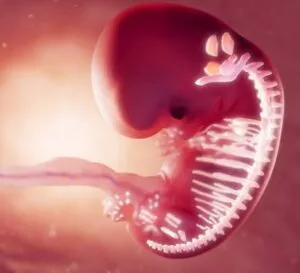
Ossification is the term used for bone formation and it continues to adulthood and beyond to repair broken bones. In addition, Osteogenesis and ossification are also terms that indicate the procedure of bone formation. Also, parts of the skeleton start forming during the very first weeks after conception. By the end of the second month, the skeletal pattern transforms into cartilage and connective tissue membranes. Accordingly, this is how ossification begins. For parents concerned about their child’s growth, our Ossification doctors at the Medical City Children’s Orthopedics and Spine Specialists Medical Practice specialize in children and their growth.
If your child needs surgery or casting, our Fracture Care Clinic opens every day and you do not need an appointment. Surgery rooms get scheduled every morning, so your child receives the care and attention they need right away.
Ossification
 Bone development continues up to the stage of adulthood. Even after adult stature fully develops, bone development continues for repairing fractures and remodeling to cope with changing lifestyles. Additionally, the three cell types involved in the development, growth, and remodeling of bones are Osteoblasts, osteocytes, and osteoclasts. Osteoblasts are bone-forming cells, while osteocytes are mature bone cells.
Osteoclasts break down and reabsorb bone. In the intricate process of endochondral ossification, these specialized cells play a crucial role by forming holes in the bone collar. This action is not just about breaking down bone, but is essential for allowing the passage of periosteal buds.
Periosteal buds consist of blood vessels, osteoprogenitor cells, and hemopoietic cells, all vital for bone development. By facilitating the entry of these components, osteoclasts contribute significantly to the transformation and growth of bone tissue. This dynamic process underscores the importance of osteoclasts in not only bone resorption but also in creating pathways for new growth and development.
The two types of ossification are intramembranous and endochondral. Quite obviously, ossification or osteogenesis plays a crucial role not only in the development and growth of bones but also in the healing of broken bones. Especially in children, broken bones have a brilliant ability to heal. New bone starts forming within a few weeks of the injury. However, complete healing tends to take longer.
What Role Does Osteoid Play in Bone Formation?
Osteoid acts as the essential foundation in the process of bone creation. Produced by osteoblasts, it forms the initial, soft organic matrix of the bone. This matrix is rich in collagen and other proteins, giving it flexibility and structure before hardening. As the osteoid accumulates, calcium salts begin to deposit within it, gradually transforming the soft matrix into rigid, mineralized bone.
Interestingly, as the bone hardens and matures, some osteoblasts become trapped within the matrix they’ve built. These cells then mature into osteocytes, which help maintain bone strength from within. The formation of osteoid is therefore a critical first step—without it, the subsequent mineralization and durability of bone tissue simply wouldn’t be possible.
QUESTIONS AND ANSWERS
How can I tell if my child has broken a bone, and what should I do if I suspect a fracture?
Common signs of a broken bone in a child include severe pain at the site of the injury, swelling, bruising, deformity, and reluctance to use the injured limb. If you suspect a fracture, it’s essential to:
- Keep the child as still as possible to prevent further injury.
- Immobilize the affected area with a splint or makeshift support, such as a rolled-up newspaper.
- Apply ice to reduce swelling and provide pain relief.
- Seek immediate medical attention, as a healthcare professional can accurately diagnose the fracture, provide treatment, and ensure proper healing.
How is a broken bone treated in children, and what is the expected recovery time?
The treatment of a broken bone in a child depends on the type and location of the fracture. Common treatments include:
- Casting: For stable fractures, our doctor will apply a cast to immobilize the bone and promote healing. Patients should wear casts for several weeks.
- Splinting: Some fractures may be treated with a splint, rather than a cast and may be used when the bone needs some mobility for proper healing.
- Surgery: In cases of complex or displaced fractures, surgery may be required to align and secure the bone with hardware, such as pins, plates, or screws.
- Recovery times vary depending on the specific fracture and the child’s age, but it can range from a few weeks to several months. Children generally heal faster than adults, but it’s essential to follow the healthcare provider’s instructions for a successful recovery.
What can I do to support my child's healing process and prevent future fractures?
To support your child’s healing and reduce the risk of future fractures:
-
-
- Ensure your child follows the prescribed treatment plan, including cast care and any physical therapy recommendations.
- Provide a balanced diet rich in calcium and vitamin D to promote bone health.
- Encourage physical activity to strengthen muscles and bones, but ensure it’s age-appropriate and safe.
- Educate your child about safety measures, such as wearing protective gear during sports and avoiding risky behaviors.
- Schedule follow-up appointments with the healthcare provider to monitor the healing progress and address any concerns.
-
It’s crucial to consult with a healthcare professional for personalized advice and guidance on your child’s specific injury and recovery process. Proper medical care and compliance with treatment recommendations are essential for a successful recovery from a broken bone.
The doctors at the Medical City Children’s Orthopedics and Spine Specialists Practice only treat children. As such, our doctors have become experts with children and adolescents and treat their broken bones. We urge parents to bring their children to us to ensure proper broken bone treatment & healing.
How Do The Broken Bones Heal?
There are three stages of bone healing: the inflammatory stage, the reparative stage, and the remodeling stage.
The Inflammatory Stage
When a bone breaks, the body sends signals to special cells to come to the injured area. Some of these cells cause inflammation in the injured area. It signals the body to stop using the injured part so that it heals better. During this stage, other cells come to the area and form a hematoma (blood clot) surrounding the broken bone. And this becomes the first bridge between the fragments of the broken bone.
Why Are Epiphyseal Growth Plates Vulnerable to Fractures?
Epiphyseal growth plates—often called “growth plates”—are the areas of cartilage near the ends of children’s bones where new bone grows. These plates are essential for healthy bone development, but are naturally softer and less dense than the surrounding mature bone. If your child sustains a significant injury, these growth plates are more likely to be damaged because they’re not as strong or rigid as fully developed bone tissue.
This structural difference makes growth plates the most common location for bone fractures in growing children. A fracture in this region isn’t just painful—it can impact the growth and shape of the bone if not treated properly. Prompt medical evaluation and specialized care are key, as injuries to the growth plate can sometimes lead to complications such as shortened or uneven limbs. This is another reason why having your child evaluated by pediatric orthopedic experts, like those at our Medical City Children’s Orthopedics and Spine Specialists Practice, is so important after any injury involving the limbs.
Closure of the Growth Plate at Puberty’s End
As puberty concludes, the growth plate—also known as the epiphyseal plate—undergoes a significant transformation. Chondrocyte activity within the plate slows down and ultimately ceases; these cartilage cells no longer proliferate. The plate itself gradually becomes entirely replaced by bone tissue, a process referred to as epiphyseal closure or ossification of the physis.
Once this ossification is complete, the capacity for bones to increase in length comes to an end. From that point forward, the only cartilage that remains is located at the joints, where it forms the articular surfaces that cushion and facilitate smooth movement throughout adulthood.
What is the Salter-Harris System, and How Does it relate to Growth Plate Fractures?
When it comes to fractures in children, understanding growth plate injuries is crucial because these areas are responsible for bone growth during childhood and adolescence. The Salter-Harris system is the most widely recognized method doctors use to classify fractures that involve the growth plate (also called the physis).
This classification system divides growth plate fractures into five main types based on where the break occurs and its pattern. Each type provides information about the injury’s severity and helps guide treatment decisions. For example:
- Type I: The fracture runs straight through the growth plate, separating the bone end from the shaft without involving the bone itself.
- Type II: The fracture passes through the growth plate and extends into the bone shaft.
- Type III: The fracture extends through part of the growth plate and into the end of the bone.
- Type IV: The break crosses both the growth plate and the bone shaft and the end.
- Type V: A compression injury damages the growth plate itself.
Correctly identifying the type of Salter-Harris fracture is essential because it helps your child’s orthopedic specialist determine the most effective treatment plan and predict the likelihood of long-term complications such as growth disturbances. Early evaluation and appropriate care are especially important at centers like ours, where specialists focus exclusively on children and teens.
The Reparative Stage
This stage starts within a week of the injury. A soft callus, a type of soft bone, replaces the blood clot that has already developed in the inflammatory stage. The callus helps hold the broken bone pieces together. However, that body part is not strong enough for use. The soft callus becomes harder over the next few weeks. And by the 2nd-6th week, this hard callus becomes strong enough to use that body part.
In the process of bone repair, the bone collar forms with three distinct yet integrated layers, each playing a crucial role:
- The outermost layer consists of osteoprogenitor cells actively dividing due to its rich blood supply. This layer becomes vital for ongoing proliferation.
- The central layer is composed of cartilage. This zone provides structural support during the repair process.
- The innermost layer features newly forming bone, replacing fragmented bone and contributing to the overall strengthening of the structure.
These layers work together seamlessly to facilitate effective bone healing and regeneration.
The Remodeling Stage
This remodeling stage starts around six weeks after the injury. During this stage, regular bone replaces the hard calluses. If you look at an X-ray of the healing bones, you will see that they look uneven. However, within the next few months, the bone retains its complete shape. And it again looks the way it did before the injury.
How Bone Remodeling Works
Bone remodeling is a sophisticated process that keeps your bones in the right shape and structure. Imagine it as a balance act between two main processes: bone resorption and bone deposition.
- Surface Remodeling: This starts the magic. Bone is deposited under specific areas of the periosteum—the outer layer of bone—while simultaneously being resorbed in other regions. This ensures that the bone adapts to various stresses and maintains its general shape.
- Endosteal Surface Activity: Inside the bone, similar activities occur. Bone is added and taken away along the endosteal surface, which lines the inner cavity of the bone. This continuous cycle allows the bone to adapt and reform as necessary.
The rate at which bone becomes deposited or resorbed directly affects whether the bone changes shape or stays the same. Over time, these processes ensure that even after an injury, your bones can return to their former glory, looking just as they did before.
Types Of Ossification
The following are the types of ossification that help in the growth and development during fetal development and whenever a bone breaks.
Intramembranous Ossification
Intramembranous ossification replaces the sheet-like connective tissue membranes with bony tissue. Bones that develop through this procedure are called intramembranous bones. This category includes certain flat bones of the skull and several irregular bones. The bones first develop as connective tissue membranes. Then, osteoblasts move to the membranes and accumulate a bony matrix around themselves. At this stage, these are called osteocytes.
In this process, mesenchymal cells play a vital role by differentiating directly into osteoblasts, which are specialized cells that secrete the bone matrix. As these osteoblasts embed themselves within the matrix they produce, they eventually transform into osteocytes. Despite the increasing distance between them, these cells remain interconnected via thin cytoplasmic processes, which are crucial for maintaining cellular communication.
As the matrix calcifies, the osteoblasts help form a network of trabeculae and spicules, establishing the bone’s internal framework. Simultaneously, surrounding mesenchymal cells differentiate into osteoprogenitor cells. These cells contact the newly formed bone spicules, transform into osteoblasts, and continue to secrete matrix, thus promoting further bone development. This ongoing growth process is known as appositional growth.
By understanding these detailed steps, we see the intricate and essential role osteoblasts play in building and shaping bones through intramembranous ossification.
The Role of Mesenchymal Cells in Intramembranous Ossification
Intramembranous ossification is a crucial process in the formation of flat and irregular bones, and the mesenchymal cells play a significant role in this mechanism.
Transformation into Osteoblasts
These versatile mesenchymal cells initially reside in connective tissue. Their primary function within intramembranous ossification is to transform directly into osteoblasts. Osteoblasts are specialized cells vital for bone formation as they are responsible for producing the bone matrix, which forms the structural foundation of bones.
Secretion and Structuring
As osteoblasts evolve from mesenchymal cells, they begin secreting the bone matrix around themselves. This secretion of matrix material is an essential phase, as it helps in the physical formation of bone.
Connectivity and Growth
Interestingly, even as the bone matrix develops, these osteoblasts do not lose contact with one another. They maintain connection through slender cytoplasmic processes, ensuring a cohesive network that supports bone growth and structural integrity.
In summary, mesenchymal cells are the foundational building blocks in intramembranous ossification, acting as the starting point from which bone-forming osteoblasts emerge, thereby creating the structure of the bone itself.
Endochondral Ossification
Endochondral ossification replaces the hyaline cartilage with bony tissues. Most of the bones develop in this way, and these bones in the skeleton refer to endochondral bones. In this procedure, the bones first develop as hyaline cartilage models. By the end of repair, endochondral ossification will convert all of the cartilage into primary bone tissue, completing the transformation efficiently.
After the third month of conception, blood vessels and osteoblasts fill the perichondrium surrounding the hyaline cartilage, which then transforms into a periosteum.
The osteoblasts create a collar of compact bone surrounding the diaphysis. This bone matrix, secreted by the osteoblasts, forms a bone collar that plays a crucial role during endochondral ossification. The bone collar acts as a barrier, preventing nutrients from reaching the hypertrophied chondrocytes, which causes them to degenerate.
Also, side by side, the cartilage in the center of the diaphysis begins to degenerate. Osteoblasts get into the degenerating cartilage and replace it with spongy bone. This develops a primary center of ossification. By understanding these interactions, we gain insight into the intricate dance of cellular activity that shapes developing bones.
Formation of the Cartilage Model
Initially, a cartilage model of the bone forms as mesenchymal cells condense and differentiate into chondrocytes, creating the hyaline cartilage model. This model sets the foundational framework for subsequent bone development.
Invasion by Blood Vessels
As the chondrocytes within this model hypertrophy, the surrounding extracellular matrix becomes calcified. Blood vessels penetrate the center, prompting the perichondrium to transform into periosteum. During this invasion, chondrogenic cells shift to osteoprogenitor cells, marking a crucial transition in bone formation.
Transformation into Bone Cells
These osteoprogenitor cells further differentiate into osteoblasts, which are pivotal in secreting bone matrix that forms the bone collar. This collar restricts nutrient access to the hypertrophied chondrocytes, leading to their degeneration and paving the way for ossification.
Role of Osteoclasts and Periosteal Buds
Osteoclasts arrive to break down sections of the bone collar, creating pathways for periosteal buds. These buds, comprising blood vessels, osteoprogenitor cells, and hemopoietic cells, are crucial for nourishing and expanding the developing bone.
Continuous Bone Development
Osteoprogenitor cells within the periosteal buds multiply, with some differentiating back into osteoblasts to continue depositing bone matrix on calcified cartilage. This synergistic process forms a calcified cartilage-calcified bone complex, ensuring the structural integrity and expansion of the bone.
Growth of the Bone Collar
The bone collar extends towards the epiphyses, while osteoclasts resorb the calcified cartilage-calcified bone complex. This action facilitates the widening of the marrow cavity, completing the development of a robust primary ossification center.
Once spongy bone grows in the diaphysis, osteoclasts break down the newly formed bone to expose the medullary cavity. Thus, the cartilage in the epiphyses continues to grow, allowing the developing bone to grow longer. Later, after birth, secondary ossification centers develop within the epiphyses. Ossification in the epiphyses mirrors that in the diaphysis, with the key distinction being that the spongy bone remains intact, not breaking down to create a medullary cavity.
Once the secondary ossification is done, the bone completely replaces the hyaline cartilage except in two areas. An area of hyaline cartilage stays over the surface of the epiphysis, known as the articular cartilage, and another area of cartilage stays between the epiphysis and diaphysis. This is the epiphyseal plate, also known as the growth region.
Secondary ossification centers play a vital role in bone growth, particularly in lengthening long bones. Located at the epiphyses, these centers undergo a process akin to primary ossification but with notable differences. Unlike primary ossification, secondary centers develop without a bone collar. Instead, osteoprogenitor cells penetrate the epiphyseal cartilage, transforming into osteoblasts and secreting matrix on the cartilage framework.
This process is essential to the bone’s ability to elongate. As the cartilage is gradually replaced by bone tissue, the length of the bone increases, allowing for growth. By understanding these mechanisms, we can appreciate how the intricate dance of cells and structures drives the development of our skeletal system.
This meticulous process ensures that by the end of the repair, the transformation from cartilage to primary bone tissue is thorough and definitive.
Bone Tissue Formation
Ossification, aka osteogenesis or bone mineralization, plays a crucial role in bone remodeling. This is the procedure of placing new bone material with the cells called osteoblasts. This procedure is also known as bone tissue formation. Two processes form normal and healthy bone tissues. However, in fracture healing, endochondral osteogenesis is the most commonly occurring process, for example, in healing fractures of long bones that are treated with plaster of Paris. On the other hand, intramembranous osteogenesis helps heal the fractures that are treated by open reduction and internal fixation with metal plates, screws, pins, rods, and nails.
Heterotopic ossification is a procedure that forms bone tissue that is often not typical, at an extraskeletal location. Calcification is often mistaken for ossification. Calcification describes the formation of calcium-based salts and crystals inside the cells and tissues. It is a procedure that happens during ossification.
Understanding the Process of Appositional Growth in Bone Formation
Appositional growth is a crucial process in bone formation that involves the thickening of bones. Here’s a breakdown of how it unfolds:
- Differentiation of Mesenchymal Cells: The process begins when surrounding mesenchymal cells evolve into osteoprogenitor cells. These are the precursors to bone-forming cells, and their transformation is essential for further development.
- Formation of Osteoblasts: Once these osteoprogenitor cells are in proximity to newly formed bone structures, they mature into osteoblasts. These are the cells responsible for new bone creation.
- Secretion of Bone Matrix: Osteoblasts secrete an extracellular matrix, which is a fundamental component of bone tissue. This matrix hardens over time as calcium deposits into it.
- Continuous Bone Addition: Through these sequential actions, the bone grows in thickness. New layers of tissue are added to existing bone surfaces, contributing to the robust structure of mature bones.
Appositional growth not only strengthens bones but also plays a vital role in their ability to adapt to mechanical stress throughout life.
The Role of a Bone Remodeling Unit in Internal Remodeling
Bone remodeling is an essential process in which the internal structure of bone adapts to various factors such as changes in weight, microfractures, and shifts in posture. This dynamic process is termed internal remodeling, and the bone remodeling unit plays a pivotal role in facilitating it.
A bone remodeling unit is composed of two main components: the cutting cone and the closing cone. These work together to maintain bone integrity and health.
Cutting Cones: The Resorption Phase
- Cutting cones are specialized structures resembling tunnels. They are created in the compact bone by cells called osteoclasts, which are tasked with breaking down bone tissue. This resorption activity is crucial for removing old or damaged bone.
- As the cutting cone enlarges, it accommodates the ingress of blood vessels, along with osteoblasts and their precursor cells, known as osteoprogenitor cells. This marks a transition point in the remodeling process.
Closing Cones: The Formation Phase
- Once the cutting cones reach their optimal size, the process of bone resorption halts. Here, the focus shifts to the closing cones.
- Osteoblasts, responsible for bone formation, begin the task of laying down new layers of bone tissue, or lamellae, encircling the blood vessels. These newly formed structures are essential for creating what are known as Haversian systems, which ensure the bone remains strong and adequately supplied with nutrients.
In essence, the bone remodeling unit is integral to the renewal and strength of bone, adapting to various physiological needs and ensuring skeletal resilience.
Understanding the Five Zones of Ossification in Bone Growth
Bone growth in length involves a complex process occurring in the epiphyseal plate and is organized into five distinct zones. Each plays a critical role in the growth and development of bones.
- Zone of Reserve Cartilage
Located the farthest from the diaphysis, this zone contains chondrocytes that are not yet actively contributing to bone growth. These cells are organized randomly and remain ready to become involved in proliferation as the need arises. - Zone of Proliferation
In this area, chondrocytes begin to rapidly divide, forming tightly packed groups. These isogenous groups are arranged in rows that run parallel to the bone’s long axis, which crucially contributes to the elongation process. - Zone of Hypertrophy
Here, chondrocytes increase in size and accumulate glycogen in their cytoplasm as they progress toward the diaphysis. As they mature, they eventually undergo programmed cell death, or apoptosis, preparing for the next stage. - Zone of Calcification
This zone sees the influx of calcium ions transported by blood vessels, which leads to the calcification of the cartilage matrix around the dying chondrocytes. Though the matrix becomes rigid, it has not yet transformed into actual bone tissue at this stage. - Zone of Ossification
Finally, osteoprogenitor cells migrate to this zone and differentiate into osteoblasts. These cells secrete bone matrix onto the calcified cartilage, marking the creation of new bone tissue as the matrix hardens into true bone.
This progression through the zones ensures the systematic growth and ossification of bone during development, highlighting the orchestrated transformation from cartilage to mature bone tissue.
Closure of the Growth Plate at Puberty’s End
As puberty concludes, the growth plate—also known as the epiphyseal plate—undergoes a significant transformation. Chondrocyte activity within the plate slows down and ultimately ceases; these cartilage cells no longer proliferate. The plate itself gradually becomes entirely replaced by bone tissue, a process referred to as epiphyseal closure or ossification of the physis.
Once this ossification is complete, the capacity for bones to increase in length comes to an end. From that point forward, the only cartilage that remains is located at the joints, where it forms the articular surfaces that cushion and facilitate smooth movement throughout adulthood.
Casts and Splints Help Broken Bones Heal
Casts and splints will help hold the broken bones in place during the healing period. After the new hard bone forms within 3–6 weeks, doctors usually remove the cast or splint. Considering the type and location of the fractured bone, our doctors may recommend various treatments:
Traditional cast
After repositioning the bone, doctors commonly immobilize the broken bone with the help of a plaster or fiberglass cast. A cast lets the bone heal in the right position. To treat fractures in the leg, foot, arm, and wrist bones, doctors often use casts.
Brace Or Functional Cast
A functional cast or brace is different from conventional cast immobilization, which allows restricted and controlled movement of nearby joints. Typically, doctors put a primary cast on the body part with the broken bone, and then, after some time, they will remove it. Thereafter, the specialists will put the limb in a functional brace, which will allow early mobility and movement.
Surgery
When the bone fracture is severe, doctors will probably need to perform surgery to fix the break. Experts expose and reposition the bone with their hands in an open reduction process. A child may require open reduction if they have complex fractures that are difficult to treat with a cast.
There Are Two Types Of Open Reduction:
Open reduction with internal fixation: This process entails attaching special metal plates or screws to the external surface of the bone. A surgeon may also set metal rods at the center of the bone to hold the fragments together in place. Open reduction with external fixation: In this procedure, surgeons place an external device on the injured limb after surgery. Specialists will place metal pins or screws below and above the fracture site to immobilize the bone while it heals and also to provide support.
When The Bone Does Not Heal Properly
In certain cases, a bone may not heal as expected if a person:
- Has a severe break
- Have the muscles, skin, and nerves been damaged in the same place as the broken bone
- Takes medicines such as corticosteroids
- Has a vitamin deficiency
- Has osteoporosis, diabetes, osteogenesis imperfecta, or anemia
- Consumes alcohol
- Smokes cigarettes
How Can I Help My Child Heal Broken Bones?
If you want to help your child recover from a broken bone, make sure that he or she:
- Follow a healthy diet with plenty of calcium and vitamin D
- Patients should take good care of their cast or splint
- Patients should follow their doctor’s directions regarding rest and/or doing any exercises
- Do not miss any of the follow-up appointments
Conclusion
Doctors consider a broken bone a common, treatable childhood injury, and therefore, do not panic if your child gets a bone injury. In most cases, breaks heal well. Moreover, kids can get back to normal life and do all their favorite activities shortly. Just make sure you consult a doctor and follow all their instructions. Selecting a pediatric orthopedic doctor for your child is a crucial decision that requires careful consideration.
By seeking recommendations, researching credentials, assessing hospital affiliations, reviewing patient testimonials, evaluating communication and bedside manner, considering the supportive team, discussing treatment options, seeking second opinions, prioritizing accessibility, and trusting your instincts, you can make an informed choice.
Remember, finding the right orthopedic doctor will ensure that your child receives the best possible care, leading to optimal outcomes and a healthier, happier future. The Medical City Children’s Orthopedics and Spine Specialists doctors only treat children. With offices in Dallas, Arlington, Flower Mound, Frisco, and McKinney, TX. Doctors Shyam Kishan, Richard Hostin, and Kathryn Wiesman have spent years studying children’s health and have devoted their lives to treating them.
____________________
Citation: National Cancer Institute – Bone Development and Growth
The medical content on this page has been carefully reviewed and approved for accuracy by the Medical City Children’s Orthopedics and Spine Specialists qualified healthcare professionals, including our board-certified physicians and Physician Assistants. Our team ensures that all information reflects the latest evidence-based practices and meets rigorous standards of medical accuracy, with oversight from our expert spine doctors to guarantee reliability for our patients.
Call 214-556-0590 to make an appointment.
Comprehensive services for children from birth through adolescence at five convenient locations: Arlington, Dallas, Flower Mound, Frisco and McKinney.